
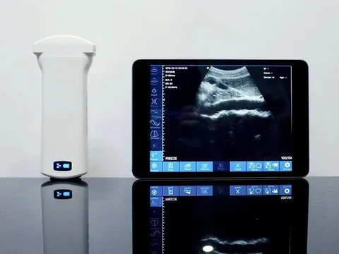
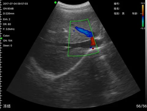
6C Convex Wireless Ultrasound Probe
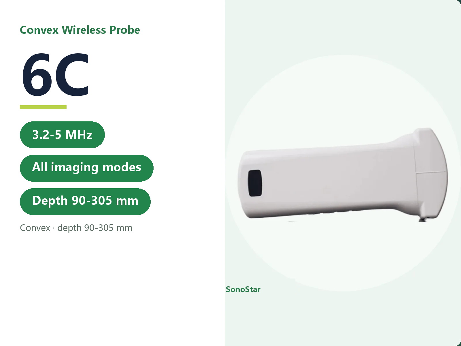
6C convex wireless probe: 3.2-5 MHz, depth 90-305 mm. Low frequency penetrates deeply for abdomen, gynaecology, obstetrics and cardiac work.
- Brand: SonoStar
- Price: On request
- Probe frequency: 3.2-5.0 MHz
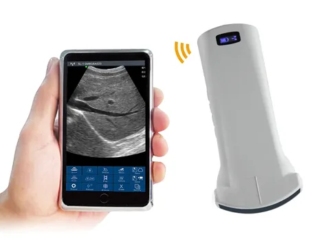
- Connection: Wireless - built-in WiFi hotspot in the probe; phone, tablet or Windows PC receives the images, with no dependence on the WiFi available on site
- Charging: Wireless charging / wired charging
- Category: Handheld Ultrasound
Product overview
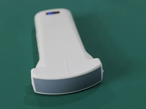
The 6C uses a convex transducer with the host built into the probe; images travel over its own WiFi to a phone, tablet or PC, so no network has to be provided on site and there is no separate main unit.
The frequency is 3.2-5.0 MHz with four depths, 90 / 160 / 220 / 305 mm. Low frequency penetrates deeply, which suits the abdomen, gynaecology, obstetrics, cardiac, urinary tract, kidney and lung.
Imaging runs in full mode - B, B/M, Color, PDI and PW - and both in-plane and out-of-plane needle guidance lines are supported, so blood flow assessment and needle guidance are both available on the same probe.
Technical specifications
Specifications for this model
| Item | Data |
|---|---|
| Probe type | Convex |
| Probe frequency | 3.2-5.0 MHz |
| Scanning depth | 90 / 160 / 220 / 305 mm |
| Imaging modes | B, B/M, Color, PDI, PW |
| Needle guidance | In-plane and out-of-plane |
| Applications | Abdomen, gynaecology, obstetrics, cardiac, urinary tract, kidney, lung |
| Colour Doppler | Steering not available; PRF 4 groups or more; wall filter 0-15; colour gain 0-100 |
| Pulsed Doppler | Sample volume 1 / 2 / 5 mm; baseline 0-800 cm/s; angle correction -85 to 85 degrees |
| Measurements | Length, angle, area/circumference, trace, area ratio; obstetric: crown-rump length, biparietal diameter, gestational sac diameter, femur length, head circumference, abdominal circumference, estimated fetal weight |
| Connection | Wireless (built-in WiFi) |
| Charging | Wireless charging / wired charging |
Common to the series
| Item | Data |
|---|---|
| Gain range | 30-105 dB |
| Noise reduction | 5 levels: 0 / 1 / 2 / 3 / 4 |
| Dynamic range | 40 / 50 / 60 / 70 / 80 / 90 / 100 / 110 |
| Battery life | About 3 hours of continuous scanning |
| Charging time | About 2-3 hours |
| Image transmission | Built-in WiFi in the probe; does not depend on the WiFi available on site |
| Devices | Phone, tablet or Windows PC (Windows software supplied) |
| Multi-screen | Android devices can mirror to a second screen over WiFi-Direct; Apple devices are not currently supported |
Specifications updated 2026-11-01
Typical applications
The application scenarios on this page describe the device based on its technical data and configuration. The actual scope of application is the one stated in the product registration certificate. Follow your institution's procedures and the instructions for use.
| Setting / department | Typical use | Configuration note |
|---|---|---|
| Emergency department | Acute abdomen, solid organ injury and rupture, fracture, internal bleeding, pleural or peritoneal fluid, pneumothorax, pericardial effusion | Convex low frequency, deep penetration |
| Intensive care | Internal bleeding, pleural or peritoneal fluid, pneumothorax and acute abdomen; pericardial effusion assessment and drainage guidance | Convex configuration, can be paired with a linear probe |
| Ambulance and pre-hospital | Organ injury, internal bleeding, pneumothorax, pleural and pericardial fluid, cardiovascular abnormality, thrombus, fracture | Convex configuration; probe WiFi independent of the site network |
| Respiratory medicine | Pleural effusion assessment and drainage guidance, alveolar-interstitial syndrome, consolidation and atelectasis | Convex configuration, can be paired with a linear probe |
| Gastroenterology | Oesophageal foreign body and disease, gastrointestinal examination (oral couplant required) | Convex low-frequency configuration |
| Hepatobiliary and general surgery | Sclerotherapy of hepatic and renal haemangioma, aspiration guidance for cysts, abscesses and pelvi-abdominal fluid; biliary puncture and organ biopsy guidance | Convex configuration |
| Urology | Percutaneous nephrolithotomy, renal biopsy, catheterisation guidance; post-void residual volume and prostate examination | Convex configuration |
| Obstetrics | Fetal development, fetal heart, cord and amniotic fluid; postpartum pelvic floor and residual volume | Convex configuration with obstetric measurement set |
Usage and selection notes
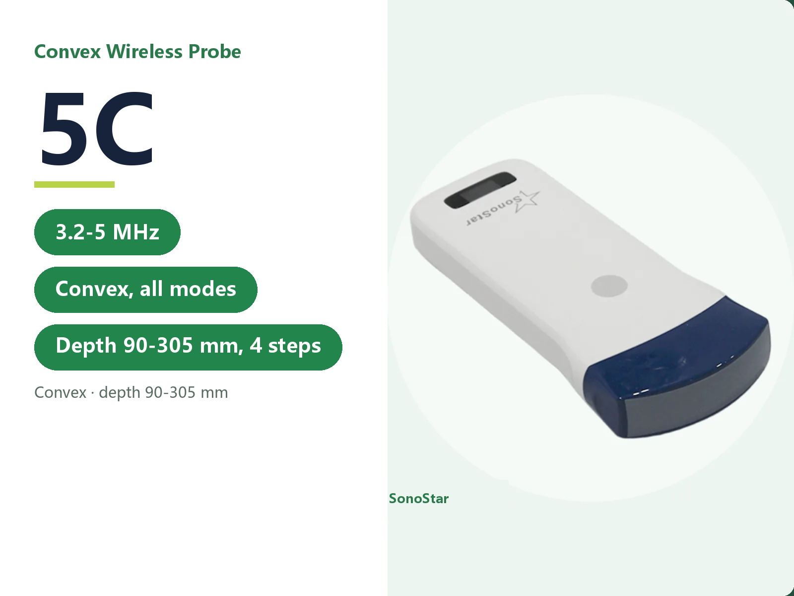
Identical in technical data to the 5C - same frequency, depths, imaging modes and applications. The two differ in configuration version and in the software they are matched to; please confirm the difference with our product department. Where superficial sites have to be covered as well, pair it with a linear probe or choose a dual-head or three-in-one model.
For contraindications and precautions, refer to the instructions for use. The device must be operated by a licensed physician with the relevant qualifications.
The technical data on this page is taken from the product technical documentation; where it has been updated, the latest version applies. The listed departments and application scenarios describe the device based on its technical data, and the actual scope of application is the one stated in the product registration certificate. This page makes no claim about clinical outcome and does not constitute clinical guidance.
Related products
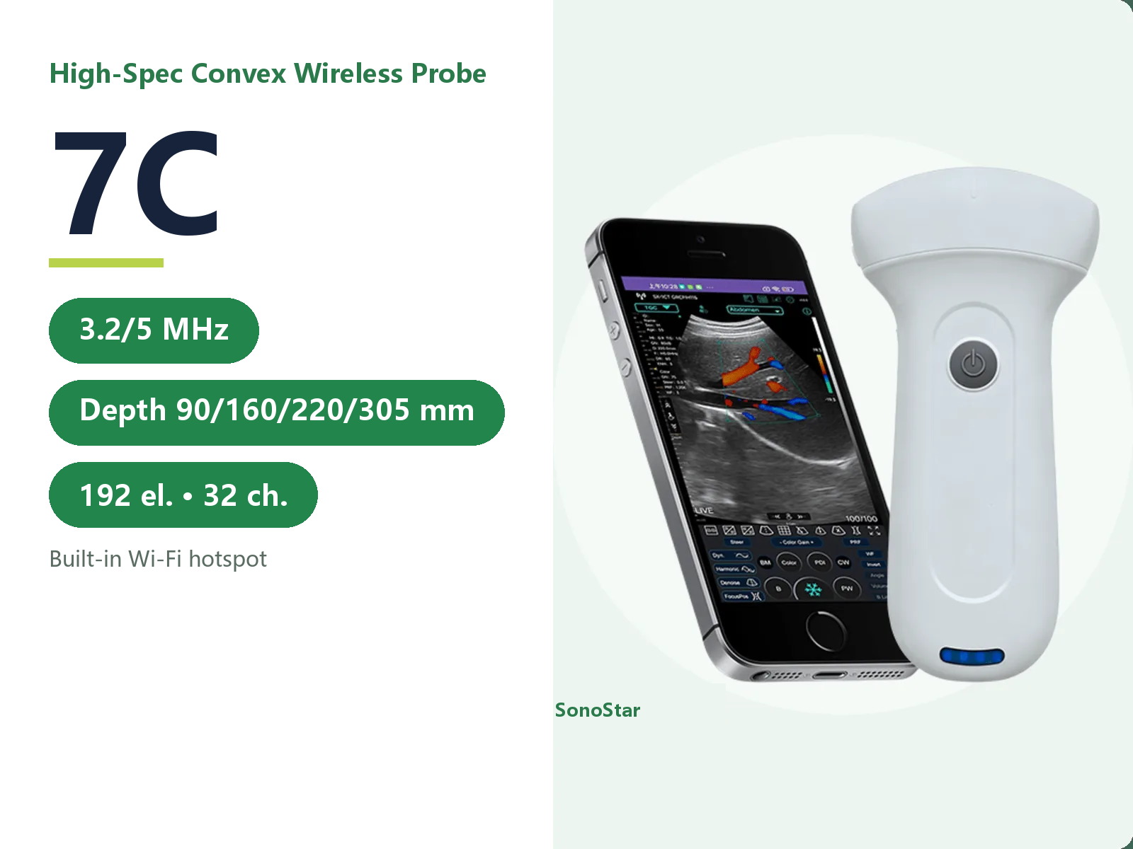
7C High-Configuration Convex Wireless Ultrasound Probe
7C convex wireless probe: 192 ...
View details →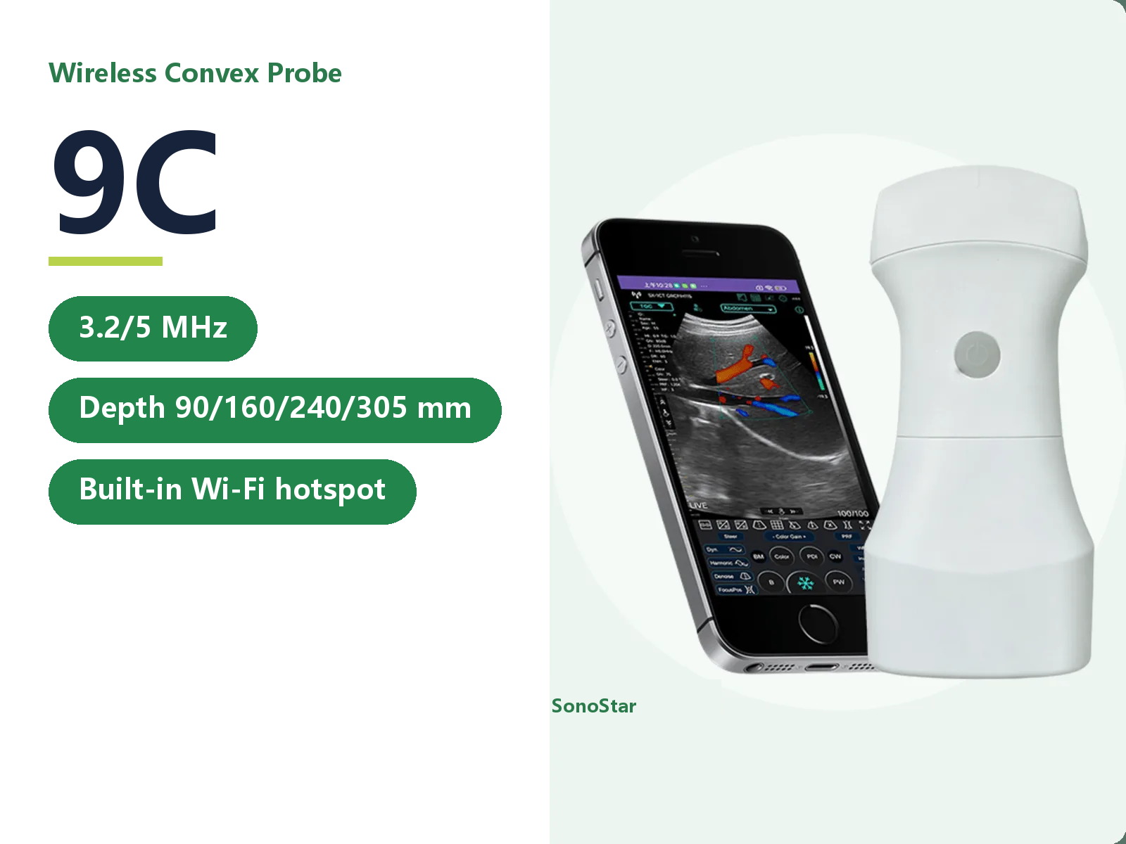
Need a quote or a sample for evaluation?
Tell us how you plan to use the system and our sales team will reply within one business day.
Get a quote
