
Ultrasound guidance in anaesthesia and pain medicine: which probes, and where the screen goes
Buying ultrasound for anaesthesia is not the same as buying it for an ultrasound department. The ultrasound department wants a system that can cover the whole body; anaesthesia wants a guidance tool that can be wheeled into the theatre and ready within a few minutes before a puncture. Most of the time it is looking at vessels and nerves a few centimetres down; occasionally at the pleura and the cardiac chambers. That pattern of use makes the priorities quite different — not bigger is better, but the closer it fits the workflow, the smoother it goes.
Three characteristics of this setting
Understand these three and a higher specification will be less likely to steer the decision.
The targets are a few centimetres down
Targets sit between the skin surface and five or six centimetres: vessels, nerve plexuses, joint spaces. A little more frequency and a little better superficial resolution are worth more here than reaching thirty centimetres.
Sterility matters more than a pretty image
A puncture is an invasive procedure. The probe has to take an isolation cover and sterile gel, and the cable and screen must not force the operator to break the sterile field — a point often overlooked and needed every day.
The theatre has no spare floor
Anaesthetic machine, monitor and drip stand already fill it. The device has to be liftable with one hand, placed at the patient's shoulder with the screen facing the operator, and out of the walkway.
Choosing the probe by guidance task
The table below maps the common tasks in anaesthesia and pain medicine to models, frequencies and penetration depths from the wireless probe range on this site; all figures come from the product specification tables.
| Guidance task | What is being visualised | Suggested probe (model / frequency / penetration depth) |
|---|---|---|
| Internal jugular and subclavian vein cannulation | Vessel course, calibre and depth | 6L, 7.5–10.0 MHz, 20 / 40 / 60 / 100 mm |
| Cervical plexus, brachial plexus and other nerve blocks | Relationship of the nerve bundle to surrounding structures | 5P, 10.0–14.0 MHz, 20 / 30 / 40 / 55 mm |
| Radial and dorsalis pedis artery cannulation | Superficial vessels, wall and lumen | 7H, 16.0–20.0 MHz, 10–40 mm adjustable |
| PICC / CVC placement | Vessel and catheter course | 5W, 7.5–10.0 MHz, 20 / 40 / 60 mm adjustable |
| Joint space and soft-tissue injection | Depth of the target and adjacent structures | 6N, 10.0–13.2 MHz, 20–60 mm adjustable |
| Lumbar puncture and epidural localisation | Interspinous space and epidural space | Choose between 5P and 6L by body habitus; the depth settings differ |
There is a point here that is easy to get backwards: higher frequency is not automatically better. High frequency gives a clear superficial image but shallow penetration; with a target five or six centimetres down in a heavier patient, the 7.5–10.0 MHz band is steadier. That is why anaesthesia departments rarely work with one probe for everything, but keep one for superficial work and one for deeper targets.
Three configurations, set by the department's main tasks
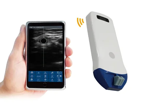
Starting point · One high-frequency superficial probe
For departments whose work is mainly central venous access, superficial nerve blocks and arterial cannulation. Representative model: the 5P high-frequency linear wireless ultrasound probe — 10.0–14.0 MHz, penetration depth 20 / 30 / 40 / 55 mm, with needle guide lines in both in-plane and out-of-plane modes.
All four depth settings sit in the superficial range, which is exactly where vessels and nerve plexuses lie. Heavier patients and deeper targets will strain it, and that is the boundary of this tier.
See the specifications →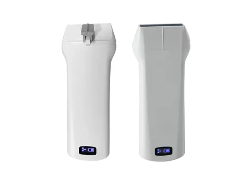
General purpose · Depth extended to 100 mm
Where body habitus varies widely and deeper targets such as the sciatic nerve are in scope, choose the 6L linear wireless ultrasound probe: 7.5–10.0 MHz, penetration depth 20 / 40 / 60 / 100 mm, again with in-plane and out-of-plane guide lines.
The trade-off in the 7.5–10.0 MHz band: superficial resolution is a little lower than above 10 MHz, but penetration is better. Most central venous access and routine nerve blocks in theatre fall inside its range.
See the specifications →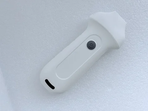
Add-on · Very high frequency for shallower layers
For musculoskeletal work, small superficial nerves, and assessment around digits and flaps, add a 7H high-specification linear wireless ultrasound probe: 16.0–20.0 MHz, penetration depth 10–40 mm adjustable.
Its value is resolution in the near field: with a target one or two centimetres down, high frequency separates the layers. Any deeper and it is out of range — which is why it is a supplementary probe rather than a main model.
See the specifications →Where a department wants one probe to cover both superficial nerve work and routine line placement, look at the 5W (7.5–10.0 MHz, depth 20 / 40 / 60 mm adjustable, wide field of view) or the 6N (10.0–13.2 MHz, depth 20–60 mm adjustable). The two cover different depth ranges; confirm against the depths the department actually works at rather than looking only at frequency.
Set it up once before buying: screen, hand and needle in relation to each other
There are no specifications in this section, yet it decides whether the device gets used at all. Take up your usual position for a puncture and try placing the device into it:
- Who the screen faces. It has to sit square in the operator's line of sight, not off to the side or behind. With one hand advancing the needle and the eyes on the screen, turning the head stops the hand.
- Where the device goes. At the patient's shoulder on the operator's side is handier than at the foot of the bed. Handheld and wireless formats can sit on the edge of the sterile trolley or on a mount; a trolley system usually means moving the bed.
- Where the cable runs. The wireless format has no cable to interfere, which is its practical advantage in theatre; with a cabled model, decide in advance which way the cable will run so that it does not cross the sterile field.
- How sterility is maintained. Probe cover, sterile gel and sterile gloves are three things, and all three are required. Before buying, confirm the probe's shape takes a standard-size cover.
- How the charge is topped up. On consecutive lists the battery is a real problem. Models with a replaceable battery (the 9L and similar) are less trouble on a full surgical day; check the specification table for the model concerned.
Three boundaries, to be written into departmental policy
- The guide line is only a reference for plane and depth. The dashed line on the screen marks the direction of the needle path; it does not mean the tip is at that position. Watch the tip throughout insertion, and stop to correct if it drifts.
- Not every patient is suitable. Infection at the puncture site, severe coagulopathy, anatomical variation, and a body habitus that prevents the target being seen clearly are all situations that call for reassessment, handled under the protocol of the institution concerned.
- It does not replace sound technique or anatomical knowledge. The device shows the structures; deciding the needle path, the depth and the risk remains with the operator. Training and supervision should come before the equipment arrives.
Common questions
Q: If the department can only have one probe, which frequency band?
A: Look at the department's main tasks. For central venous access and routine nerve blocks, the 7.5–10.0 MHz band covers more ground; for superficial nerves and arterial cannulation, above 10 MHz suits better. If unsure, tally a month of task types and decide by proportion.
Q: Will a wireless probe be interfered with in theatre?
A: The wireless models emit their own WiFi signal from the probe, received directly by a phone, tablet or Windows computer; they do not depend on the theatre having a network. Keep the receiving device close and avoid thick lead doors in between.
Q: Do we need both in-plane and out-of-plane guidance?
A: Most of the linear probes on this site support both modes; check the needle guide line row of the specification table for the model concerned. In-plane shows the full length of the needle, out-of-plane shows only its cross-section. They suit different situations, and in daily work both get used.
Q: On consecutive lists, will the battery cope?
A: Estimate from the battery figures in the product specification table against the surgical schedule. In practice departments usually keep a second probe spare, or choose a model with a replaceable battery, and recharge between cases rather than during one.
Q: Can it be used for preoperative gastric content assessment?
A: That is a clinical assessment task requiring its own training and departmental protocol, and it is not something equipment configuration can answer. What can be confirmed at the device level is whether the depth settings of the convex and linear probes cover the region you need to see — check against the specification table for the model.
Related reading
Ultrasound guidance in anaesthesia: how to configure the equipment and choose the probe
The full treatment of the same subject, covering four categories of guidance task and the details of sterile technique.
Read →Ultrasound-guided nerve block: the full sequence from finding the nerve to injecting
The order of steps from locating to injecting, plus a pre-puncture checklist.
Read →In-plane or out-of-plane: choosing between the two approaches
What each needle plane shows, and what it does not.
Read →Ultrasound-guided central venous access: approaching the internal jugular and the subclavian
Position, view and points to watch on each route.
Read →List the tasks first, then choose the model by depth
The Selection Guide section of this site carries a wireless probe comparison table that puts model, frequency and penetration depth on a single sheet, so anaesthesia and pain departments can check it task by task against their own workload.
Download the selection guideThe specifications in this article are taken from the product specification tables on this site. This document is a reference explanation based on technical parameters; where it differs, the content stated on the product registration certificate takes precedence. For contraindications and precautions, see the product instructions for use. The product must be operated by a licensed physician with the appropriate qualifications.

