
Urology ultrasound: separate how much urine from what it looks like
The two questions that come up most often in a urology clinic are, at root, two different things. One is quantitative — how much urine is left in the bladder, whether to catheterise, and how much to drain. The other is about pictures — is there hydronephrosis, what does the bladder wall look like, how large is the prostate. The first is answered by a single probe measuring volume directly; the second needs a convex probe for deep structures, an endocavity probe for the transrectal route and a linear probe for vascular access. Discuss all four as one equipment decision and the configuration can never be settled.
Four constraints to work through first
Decide first whether you need a number or an image; the rest of the choice becomes much clearer.
Most often what is wanted is a number
Post-void residual is the most common objective measurement in urology. What it needs is reproducible volume measurement, not an attractive image — and those two things ask different things of a device.
Deep structures need low frequency
Kidneys, bladder and renal pelvis all sit deep in the abdomen, more so in obese patients. The convex 3.2–5.0 MHz band reaching 305 mm is the baseline for structural work.
The prostate needs a short route
The transrectal route puts the transducer beside the gland: endocavity probe, 6.0–8.0 MHz, 30–120 mm depth. Transabdominal views of the prostate fight both distance and bowel gas.
Dialysis access is a separate matter
Fistulas, grafts, thrombus and stenosis are superficial vascular work needing a high-frequency linear array. That is not the same probe band as abdominal structural examination.
Which category of equipment each routine urology examination falls into
The models and figures below come from the specification tables on this site and can be checked item by item against the product pages.
| Question | Equipment and probe | Frequency / depth |
|---|---|---|
| Post-void residual; whether catheterisation is needed | Bladder scanner | 2.6 / 5 MHz four-dimensional scanning, volume measured directly |
| Visualising catheterisation (avoiding blind insertion) | Bladder scanner | 2.6 / 5 MHz image while draining, probe suprapubic |
| Kidney and bladder structure, calculi | Convex | 3.2–5.0 MHz 90 / 160 / 220 / 305 mm |
| Prostate (transrectal) | Endocavity | 6.0–8.0 MHz 30 / 60 / 90 / 120 mm |
| Prostate (transrectal, close-range setting) | Endocavity | 6.6–8.0 MHz 20–100 mm |
| Dialysis access, fistula, thrombus and stenosis | Linear | 10.0–13.2 MHz 20–60 mm |
| Vascular puncture and line placement guidance | Linear | 7.5–10.0 MHz 20–100 mm |
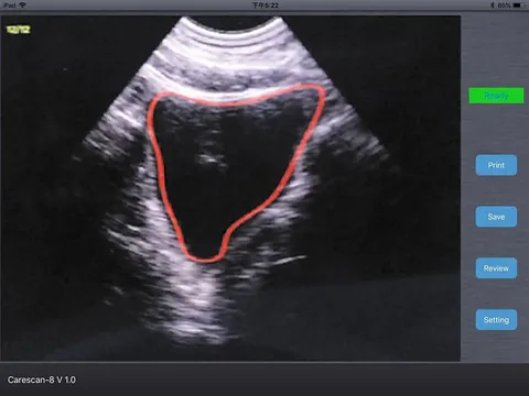
The first two rows deserve separate comment. A bladder scanner is a single probe that sends the image wirelessly to a phone or tablet and measures volume by four-dimensional scanning. According to the product instructions it measures post-void residual, on which basis the decision is made whether to catheterise and how much volume it is appropriate to drain; during catheterisation it provides visual monitoring so that the catheter is not inserted blind and does not cause injury. In other words, what it provides is the volume and the timing of drainage — not an imaging diagnosis of the bladder wall or the shape of the prostate.
The third and fourth rows are purely about looking: the convex probe covers overall kidney and bladder structure, the endocavity probe covers the prostate transrectally. Where both are routine, the least troublesome choice is a model with two active faces — one device, switched by button, with no changing of machines.
Four common configurations, chosen by what the department actually does
From solving urine volume alone to covering the whole department's structure and access work, these are four different boundaries.
Volume only · One probe
For residual urine screening on wards, in nursing units and rehabilitation, or for judging the timing of catheterisation in a urology clinic. Representative models: the BProbe-2 wireless bladder scanner (2.6 MHz) and the BProbe-5 (5 MHz) — both use four-dimensional scanning, and the probe itself is the entire device, with no mainframe needed.
The trade-off is direct: it gives no structural detail of the bladder wall, prostate or kidneys, only volume and a guide to catheterisation. Structural examination in clinic requires separate ultrasound.
See the specifications →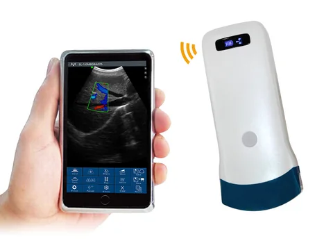
Volume plus structure · Adding one convex probe
For a routine urology clinic that needs kidneys, bladder, calculi and residual urine together. Alongside the scanner, add a 5C convex wireless ultrasound probe: 3.2–5.0 MHz, penetration depth 90 / 160 / 220 / 305 mm. The urological uses listed in the product instructions include post-void residual, prostate examination and guidance for catheter insertion.
This is where most urology departments in secondary hospitals start: one probe covers abdominal structure, and residual urine can go by either route.
See the specifications →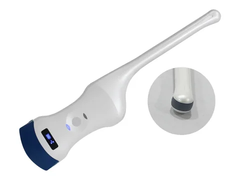
Adding the transrectal route · One device covers both
Where prostate examination is routine. Representative model: the 5CT convex plus endocavity wireless ultrasound probe — convex 3.2–5.0 MHz, depth 90 / 160 / 220 / 305 mm; endocavity 6.0–8.0 MHz, depth 30 / 60 / 90 / 120 mm. The urological uses listed in the product instructions are post-void residual, prostate examination and guidance for catheter insertion.
One probe, two active faces: convex for kidney and bladder, endocavity for the transrectal prostate. The endocavity face needs covers and a fixed cleaning routine.
See the specifications →The fourth is a tier kept for dialysis and vascular access. The 6N high-specification linear probe (10.0–13.2 MHz, depth 20–60 mm) or the 7L high-specification linear wireless ultrasound probe (7.5–10.0 MHz, depth 20–100 mm) both list vascular access, thrombus and stenosis assessment, and puncture guidance among their uses, and can be applied to fistulas, grafts and line assessment. This band reaches only tens of millimetres, which is a quite different matter from abdominal structural examination — do not try to make a convex probe cover it.
Where volume is high and deeper abdominal scanning is needed, move up within the convex tier: the 7C (3.2–5.0 MHz, depth 90 / 160 / 220 / 305 mm, 192 elements) or the 8C (3.2–5.0 MHz, depth 90 / 160 / 240 / 300 mm). The depth settings differ; confirm against actual body habitus and examination depth.
Four things that often get missed
- Residual urine is an indirect measure. In the same patient the value can differ with position, time and voiding history. So the bladder should be emptied beforehand, the position kept consistent, and the conditions written into the record — a single value is not a basis for judgement; the trend is what carries meaning.
- A scanner measures volume, not the wall. It can answer how much urine there is; it cannot answer whether the bladder wall is thickened or whether there is a prostatic nodule. Separating the two in the departmental protocol prevents the misuse of treating a volume reading as an ultrasound examination.
- Consumables and workflow for the transrectal route. The endocavity probe contacts mucosa, so single-use covers, sterile gel and post-examination cleaning have to be fixed as a routine. That cost and time belongs in purchasing and scheduling decisions; the device price alone is not the whole sum.
- Access assessment for dialysis patients has to be scheduled separately. Fistula, graft, thrombus and stenosis assessment needs a high-frequency linear probe and a consistent position; it is a different set of movements from routine abdominal scanning. A department with dialysis work is better served by listing this item separately and assigning equipment and time to it than by borrowing a device as needed.
Three lines not to cross
This section matters more than every specification above it.
- Residual volume alone is not a basis for management. Whether to catheterise, how much to drain, whether to leave a catheter in place — these need clinical findings, renal function and voiding history taken together. The device provides a measurement; the decision belongs to the physician. The indications and contraindications for catheterisation in the instructions for use and in institutional policy take precedence over any device reading.
- Judging how enlarged the prostate is, is not the same as grading it. Ultrasound shows gland size and shape, but symptom scoring, uroflowmetry, PSA and biopsy where indicated are separate steps. Replacing that assessment pathway with ultrasound alone is a common misunderstanding.
- The transrectal route has indications and contraindications. Not every patient is suitable for it. The specific indications, contraindications and preparation follow institutional policy and the instructions for use, and are outside the scope of this page.
One more point worth stating up front: imaging findings in urology are usually read together with urodynamics, laboratory tests and endoscopic appearances. The structure ultrasound shows and the symptoms the patient reports are two parallel lines of information, and neither replaces the other.
Common questions
Q: Can a bladder scanner replace conventional ultrasound?
A: No — it solves a different problem. A scanner measures volume and gives a reference for the timing of catheterisation, with one probe connecting wirelessly to a phone or tablet. Conventional ultrasound looks at kidney, bladder wall and prostate. Where structural information is needed, a convex or endocavity probe is still required.
Q: What residual volume counts as abnormal?
A: The threshold is set by the department according to the guideline it follows and the individual patient; the device's job is to measure the volume accurately. One reminder: values fluctuate under different conditions in the same patient, so record whether the bladder was emptied beforehand, the position and the time of measurement, and compare on the same terms at follow-up.
Q: What does transrectal prostate examination require?
A: An endocavity probe in the 6.0–8.0 MHz, 30–120 mm band, single-use covers and a fixed cleaning routine, plus indication assessment and patient preparation in line with institutional policy. The transabdominal route can also show the prostate, but distance and bowel gas work against it, and detail is poorer than transrectal.
Q: Which probe for access assessment in dialysis patients?
A: A high-frequency linear array. The 6N (10.0–13.2 MHz, depth 20–60 mm) suits superficial access detail; the 7L (7.5–10.0 MHz, depth 20–100 mm) reaches a little deeper. Both list vascular access, thrombus and stenosis assessment and puncture guidance among their uses.
Q: Can one device cover kidney, bladder and prostate?
A: It has to carry two active faces. Convex covers kidney and bladder, endocavity covers the transrectal prostate; the 5CT (convex 3.2–5.0 MHz plus endocavity 6.0–8.0 MHz) puts both on one probe, switched by button. A convex-only probe cannot do the transrectal prostate, and an endocavity-only probe cannot see the kidney.
Related reading
Handheld, portable or trolley ultrasound: start with where it will sit
Moving between clinic and ward, form factor often decides usability more than specifications do.
Read →Ultrasound-guided versus landmark puncture: where each approach fits
Line placement, stoma formation, puncture guidance — what being able to see changes.
Read →Obstetrics and gynaecology: assessing the postpartum pelvic floor and residual urine
The same residual urine question is used somewhat differently in obstetrics and gynaecology.
See the scenario →Bladder scanners: a model-by-model comparison
The difference between the two BProbe probes can be compared directly in the specification tables.
See the product section →Work out which examinations are frequent and which are occasional
Residual urine, prostate, calculi and dialysis access do not share the same equipment boundary. The Selection Guide section of this site carries a comparison table arranged by examination site and penetration depth, for discussion within the department.
Download the selection guide See other scenarios →The specifications in this article are taken from the product specification tables on this site. This document is a reference explanation based on technical parameters; where it differs, the content stated on the product registration certificate takes precedence. For contraindications and precautions, see the product instructions for use. The product must be operated by a licensed physician with the appropriate qualifications.

