
Bedside ultrasound in the emergency department: four requirements for the device, and three lines not to cross
Using ultrasound in the emergency department starts with accepting something counter-intuitive: it delivers a rapid judgement at the bedside, not a final diagnosis. Who uses it well often has less to do with how high the specifications are stacked than with two plainer things — how many seconds pass from trolley to power-on, and whether the device can be brought to the bedside at all — and with whether the operator knows what an inconclusive image looks like. This page follows the actual flow of an emergency department to set out how to configure the equipment and where to draw the boundaries.
Four tests any emergency device has to pass first
The order matters: whether it can be used comes before how well it images.
It has to start fast
Nobody wants to wait for a self-test during a resuscitation. The time from switching on to first image decides whether the device gets used at all.
It has to reach the bedside
Emergency patients are not in the ultrasound department. A trolley takes up space and will not clear a corridor corner; portable and handheld formats have a natural advantage here.
One question at a time
Is there pericardial fluid? Is there free fluid in the abdomen? Is there a pneumothorax? Choose the view by the question, not by the idea of running a complete examination.
Power and network cannot be preconditions
Sockets are in short supply in a resuscitation room, and an ambulance has no network. A wireless probe that carries its own WiFi works as normal in both places.
The questions asked at the bedside, and which probe answers them
Bedside targets in the emergency department cluster around three questions — is there fluid, is there free gas, is the vessel open — and the depth range runs from 2 cm to 30 cm.
| Question at the bedside | Probe type | Frequency | Penetration depth |
|---|---|---|---|
| Free abdominal fluid; liver, gallbladder, pancreas, spleen, kidneys | Convex | 3.2–5.0 MHz | 90 / 160 / 220 / 305 mm |
| Pleural effusion, lung sliding | Convex | 3.2–5.0 MHz | 90 / 160 / 220 / 305 mm |
| Pericardial effusion, chamber proportions | Phased array | 2.2–3.6 MHz | 90 / 160 / 200 / 240 mm |
| Pneumothorax (pleural line and lung sliding) | Linear | 10.0–12.0 MHz | 20 / 40 / 60 / 80 mm |
| Internal jugular and subclavian vein cannulation | Linear | 10.0–12.0 MHz | 20 / 40 / 60 / 80 mm |
| Rib and long-bone fracture screening | Linear | 10.0–12.0 MHz | 20 / 40 / 60 / 80 mm |
The frequencies and depths in this table come from the wireless probe specification tables on this site. A pattern shows up: abdomen and chest need low frequency and deep penetration, while pneumothorax, vessels and fractures — all superficial targets — want a higher frequency instead. That is why an emergency department rarely equips itself with a single probe.
Three common configurations, chosen by the department's flow
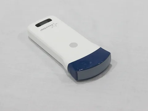
Single probe carried on the person · Abdomen and chest only
For pre-hospital use, ambulances and emergency departments staffed by a single clinician. Representative model: the 5C convex wireless ultrasound probe — 3.2–5.0 MHz, penetration depth 90 / 160 / 220 / 305 mm. The probe carries its own WiFi and does not depend on a network being present.
One probe covers the three is-there-fluid questions — abdomen, chest and pericardium. The trade-off is explicit: superficial vessel work and the detail of lung sliding are given up.
See the specifications →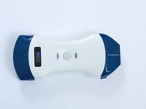
Dual head · Adding the superficial half
For resuscitation rooms and observation units that need abdominal views as well as vascular access and pneumothorax assessment. Representative model: the 5CP dual-head wireless ultrasound probe — convex 3.2–5.0 MHz, depth 90 / 160 / 220 / 305 mm; linear 10.0–12.0 MHz, depth 20 / 40 / 60 / 80 mm.
One probe, two active faces, and no swapping back and forth during a resuscitation. For most secondary hospitals this is the practical dividing line.
See the specifications →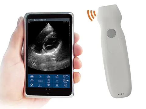
Adding a dedicated cardiac probe
The heart wants low frequency and a sector shape, which a convex array cannot force itself into. Representative model: the 5PA phased-array wireless ultrasound probe — 2.2–3.6 MHz, penetration depth 90 / 160 / 200 / 240 mm, 128 elements and 32 channels.
It is for rapid assessment of pericardial fluid, chamber proportions and volume status. Quantitative cardiac measurement remains with the ultrasound department, and that should be written into the departmental protocol.
See the specifications →Where the emergency department and the ICU share one set of equipment, look at the 6C (3.2–5.0 MHz, depth 90 / 160 / 220 / 305 mm) or the 8C (3.2–5.0 MHz, depth 90 / 160 / 240 / 300 mm). The depth settings differ between the two; confirm against the depths the department actually needs rather than guessing from the model number.
Three lines not to cross
This section matters more than every specification above it.
- A negative single scan does not exclude injury. Free fluid has a volume threshold and may not be visible early; bowel gas and subcutaneous emphysema obscure the view. A bedside result has to be read alongside history, signs and other investigations — an image alone cannot support a conclusion that nothing is wrong.
- A limited window has to be recorded honestly. Body habitus, position and gas all degrade the image. Writing "inadequate visualisation" is far more accurate than writing "no abnormality seen", and the two carry quite different weight if the case is reviewed later.
- It does not replace a formal examination in the ultrasound department. What the bedside study answers is whether something is there and roughly where; organ detail, flow assessment and quantitative measurement still go to ultrasound. Writing this into the working agreement between the two departments is more useful than putting it on a wall.
One thing to add: image quality depends heavily on the operator's experience. On the same device, a trained and an untrained user produce conclusions of different value. That should be said plainly to the department before purchase — the device only makes seeing possible; the judgement stays with the person.
Common questions
Q: Handheld wireless probe or trolley system — how should an emergency department choose?
A: Start by asking where the device will mainly be used. Moved between resuscitation room, observation unit and ambulance, a handheld or portable format is the better fit, because it gets into the room and onto the vehicle; fixed in an emergency ultrasound room and scanning dozens of patients a day, a trolley's screen and control panel are less tiring. The two divide the work by setting; they do not replace each other.
Q: Is the wireless connection unstable?
A: On the wireless models the probe itself emits the WiFi signal and the phone or tablet connects to it directly; the traffic never passes through the building's network. The things to watch are distance between the receiving device and the probe, and thick metal in between. In ordinary bedside use there is no problem.
Q: Will the battery last a shift?
A: Battery figures for the wireless probe series on this site are given in the product specification tables and vary with how hard the device is worked. In practice emergency departments usually keep two units in rotation, or choose a model with a replaceable battery, so that a busy shift does not depend on a charge gauge.
Q: Can one convex probe also image the heart?
A: A convex array can reach the cardiac region, but the sector field and low-frequency penetration of a phased array suit chamber anatomy better. Where pericardial fluid and chamber proportions are assessed routinely, add a phased array rather than making the convex array do the job.
Q: How are images archived?
A: Handheld and wireless models generally store images and clips on the phone or tablet, then export them for archiving in line with institutional rules. For the bedside record, note the time, the view, the findings and any reason the window was limited — in review these are consulted more often than the images themselves.
Related reading
Bedside ultrasound in emergency care: what the device has to do, and what must be said up front
Equipment and protocols seen from the angle of interdepartmental working — a complement to this page.
Read →Ultrasound-guided versus landmark puncture: where each approach fits
Vascular access is routine in the emergency department; this page sets out what being able to see changes.
Read →Ultrasound-guided central venous access: approaching the internal jugular and the subclavian
How the two routes differ, and the choice of position and view.
Read →Handheld, portable or trolley ultrasound: start with where it will sit
Form factor before specifications — nowhere more so than in emergency care.
Read →Match the specifications to the views, then talk about configuration
The Selection Guide section of this site carries a wireless probe comparison table, listing models by examination site and penetration depth. It can support equipment discussions in emergency departments and ICUs.
Download the selection guideThe specifications in this article are taken from the product specification tables on this site. This document is a reference explanation based on technical parameters; where it differs, the content stated on the product registration certificate takes precedence. For contraindications and precautions, see the product instructions for use. The product must be operated by a licensed physician with the appropriate qualifications.

