
Musculoskeletal ultrasound: use the depth settings to narrow the models to three
Musculoskeletal work is one of the few areas of ultrasound where shallower is harder. The tendons, ligaments, nerves and bursae to be examined mostly lie between 10 and 40 mm below the skin: too low a frequency and the layers blur together, too high and the beam will not penetrate. So the right order in musculoskeletal selection is not to pick a model but to establish how deep the structures actually are, then match frequency and depth setting to that. The linear probes in the range on this site run from 18.0–24.0 MHz down to 7.5–10.0 MHz, which divides neatly into three depth bands — and this page is that division.
Four constraints to work through first
The order matters: establish how deep, then discuss how much detail.
The targets are almost all superficial
Tendons, ligaments, bursae and subcutaneous tissue nearly all fall within 40 mm. That means high frequency is needed here rather than wasted — the 16.0–24.0 MHz band exists for exactly that thickness. The product pages on this site describe this class of probe in a consistent sentence: frequency above a conventional linear array, shallower penetration, suited to close observation of fine superficial structures.
But the thickness varies enormously
A flexor tendon in a finger and the muscle belly of a thigh differ in depth by three or four times. The same 18.0–24.0 MHz probe that reaches 25 mm is not enough on a thigh; go the other way and a probe covering 20–60 mm is not fine enough in the first few millimetres on a finger. That is why musculoskeletal services rarely manage with one probe.
What it is compared with is last time
In musculoskeletal work, follow-up and comparison with the contralateral side are routine. That requires the same view and the same depth setting on both occasions. The device supplies measurement tools and depth steps; comparability comes from protocol — and that matters more than any line in a specification table.
One hand has to be able to press the probe down
Musculoskeletal examination is a matter of technique: the probe has to be pressed, angled and slid along the long axis of a tendon. The weight and thickness of the probe directly affect whether it can be held steady for any length of time. The linear probes in this range are mostly 180–250 g at 140–156 mm, designed from the outset for one-handed use.
Cut into three depth bands and the models select themselves
The frequencies, depths and element counts below come from the specification tables for the 8S, 8H, 7H, 5W and 5P on this site and can be checked item by item.
| Layer to be examined | Model · frequency | Scanning depth settings |
|---|---|---|
| Skin, subcutaneous tissue, the most superficial bursae | 8S · 18.0–24.0 MHz | 10 / 15 / 20 / 25 mm |
| Small joints and superficial tendons of fingers and toes | 8S · 18.0–24.0 MHz | 10 / 15 / 20 / 25 mm |
| Tendons and ligaments around wrist, elbow and ankle | 8H · 16.0–20.0 MHz | 10 / 20 / 30 / 40 mm |
| Thicker muscle bellies, deeper tendons | 7H · 16.0–20.0 MHz | 10–40 mm adjustable |
| Thigh, buttock and other thick regions | 5W · 7.5–10.0 MHz | 20 / 40 / 60 mm |
| Superficial nerves and vessels | 5P · 10.0–14.0 MHz | 20 / 30 / 40 / 55 mm |
| Where a whole long structure has to be seen at once | 5W · face width 80 mm | 20 / 40 / 60 mm |
What should be copied out of this table is the depth settings, not the model names. The four 8S settings are 10 / 15 / 20 / 25 mm, the 8H has 10 / 20 / 30 / 40 mm, the 5W has 20 / 40 / 60 mm and the 5P has 20 / 30 / 40 / 55 mm. Write down the depth of the structure you need to see, find which set contains it, and the range of models is settled.
One more line is easily missed: the 5W's face is 80 mm wide, while the scanning widths of the 7H, 8H and 8S are 18 mm, 18 mm and 15 mm. A fourfold difference in width changes the way the probe is used entirely — a narrow face belongs pressed on one point for close observation, a wide one belongs slid along the long axis to take in a whole tendon or muscle in one pass. The 5W's specification table gives 7.5–10.0 MHz, an ordinary linear band; its strength is the 80 mm field and the 256 elements, not a higher frequency.
Element count and channel count are worth a look too. The 7H has 192 elements and 32 channels; the 8H and 8S have 192 elements and 64 channels; the 5W has 256 elements and 32 channels. Element count decides the fineness of the beam, channel count affects parallel processing capacity. They are not the same measure, so do not merge them into a single higher-is-better reading of the specification table.
Three common configurations, chosen by how shallow the work is
The three tiers correspond to three depth ranges; they are not a cheaper-to-better ladder.
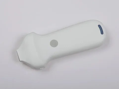
Shallowest tier · 8S
For structures within 25 mm only: skin, subcutaneous tissue, the small joints and superficial tendons of fingers and toes. Representative model: the 8S ultra-high-frequency linear wireless probe — 18.0–24.0 MHz, depth 10 / 15 / 20 / 25 mm, 192 elements, 64 channels, scanning width 15 mm.
The trade-off is plain: beyond 25 mm nothing comes back. Its value is the resolution within those 25 mm; past that depth the probe should be changed, and forcing the gain up only amplifies the noise along with the signal.
See the specifications →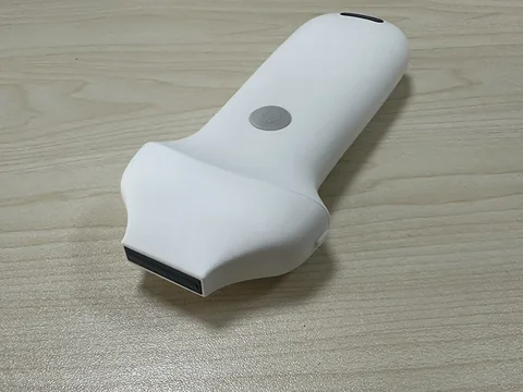
Main tier · 8H
16.0–20.0 MHz with depths of 10 / 20 / 30 / 40 mm, covering the great majority of tendons and ligaments. Representative model: the 8H ultra-high-frequency linear wireless probe — 192 elements, 64 channels, scanning width 18 mm, colour steering angles of −12 / −7 / 0 / 7 / 12 degrees, pulsed-wave Doppler correction angles from −85 to 85 degrees, and sample volumes of 1 / 2 / 5 mm.
Why this tier is called the main one: 16.0–20.0 MHz sits in the band that both separates the layers and still penetrates to 40 mm, and 40 mm already covers most tendons and ligaments of the limbs. What the 8H adds over the 7H is 64 channels and a fully listed set of colour and Doppler parameters.
See the specifications →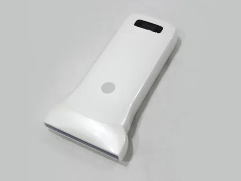
Deeper, thicker regions · 5W
For thick regions such as thigh and buttock, or where a whole long structure has to be taken in at once. Representative model: the 5W ultra-wide linear wireless probe — 7.5–10.0 MHz, depth 20 / 40 / 60 mm, 256 elements, 32 channels, a face 80 mm wide, needle-tip enhancement and 2.5 hours of battery.
The trade-off: the 80 mm field buys a wider stretch per pass, at the cost of the ends lifting when the probe is placed on a small joint. It suits one sweep along the long axis, not working over a single point for detail.
See the specifications →One more deserves separate mention: the 5P high-frequency linear wireless probe, 10.0–14.0 MHz with depths of 20 / 30 / 40 / 55 mm. Its band sits between the 8H and the 5W, and what really differs is the measurement set — length, angle, area/circumference, trace and area ratio, all five listed in the specification table. Where musculoskeletal follow-up compares the size of the same structure over time, that matters more than frequency.
Where musculoskeletal volume is modest but superficial and abdominal work are both needed, there is a step back available: a 10.0–12.0 MHz dual-head probe such as the 5MN covers superficial and abdominal work, accepting that detail shallower than 40 mm will not match a dedicated high-frequency probe. That is a trade-off, not a compromise.
Four things that often get missed
- Fix the depth setting. Examine the same structure twice at different settings and the magnification differs, so it looks as though it has changed. The remedy is plain: write one setting per commonly examined site into the departmental protocol, and note it in the report.
- Probe pressure changes the image. Musculoskeletal work requires pressure, and firm pressure compresses the superficial tissue so depth readings shift with it. This is not a device problem but a technique problem; the remedy is to standardise the pressure and state the conditions in the report.
- Gel and probe covers affect the near field. A thick layer of gel, or imaging through a single-use cover, blurs the first few millimetres. To use the 18.0–24.0 MHz band on subcutaneous tissue, those details have to be dealt with first.
- Battery life does not match continuous scanning. The 7H is rated at 1.5 hours; the 8H and 8S at 1.5 hours, or 3 with the extended battery; the 5W at 2.5 hours; the 5P's specification table does not list battery life separately. A full morning in clinic needs a charging window in between, or the extended-battery version outright.
Four lines not to cross
This section matters more than every specification above it.
- Higher frequency does not mean better. 18.0–24.0 MHz covers only 25 mm, and beyond that depth the image degrades quickly. Selling high frequency as the headline claim is the one thing most likely to be questioned on a return visit.
- Models do not map one-to-one onto body sites. The product pages for these linear probes carry the same note: the frequency is close to other models in the series and the depth settings differ slightly, so confirm the specific selection with our product team. In other words, the official position is to choose by depth, not that a particular model is for a particular body part.
- Imaging findings are not diagnostic conclusions. Continuity of a tendon, thickness of a ligament, fluid in a bursa are all imaging descriptions; whether there is a tear or inflammation is judged with history, physical examination and other investigations. The device supplies images and measurements; the judgement belongs to the physician.
- Therapeutic procedures are outside this page. Injections and release procedures common in musculoskeletal practice are clinical management, a separate matter from equipment selection. This page deals only with what can be seen and measured.
One further point: examination standards, positioning and view criteria for musculoskeletal ultrasound are set by professional bodies and by the institution concerned. What this page gives is a device-side mapping of frequency to depth, and it does not replace any protocol.
Common questions
Q: Why does musculoskeletal work need frequencies above 16 MHz?
A: Because the targets are superficial. Tendons, ligaments and subcutaneous tissue mostly lie within 40 mm, and higher frequency means a shorter wavelength, so more layers can be separated at the same depth. The cost is penetration: the 8S at 18.0–24.0 MHz reaches only 25 mm, the 8H at 16.0–20.0 MHz reaches 40 mm. Frequency and depth are a pair that must be read together.
Q: What is the difference between the 8H and the 7H?
A: The frequency is the same (16.0–20.0 MHz); the depth is presented differently. The 7H is continuously adjustable from 10–40 mm with 192 elements and 32 channels; the 8H has four settings at 10 / 20 / 30 / 40 mm, 192 elements and 64 channels, and lists colour steering angles (−12 to 12 degrees), pulsed-wave Doppler correction angles (−85 to 85 degrees) and sample volumes (1 / 2 / 5 mm) item by item. For finer colour and Doppler adjustment, choose the 8H.
Q: What is the 80 mm face on the 5W for?
A: Covering a longer stretch in one pass. The 7H and 8H scan 18 mm wide and the 8S 15 mm, so a whole tendon along its axis takes several passes; the 5W's 80 mm takes it in one. The cost is that on a small joint or a hollow the ends lift and the near field suffers. It is a tool for sweeping long structures, not for working over detail.
Q: Is needle-tip enhancement needed?
A: It depends on whether puncture guidance is part of your work. Among these linear probes, the 5W specification table explicitly lists needle-tip enhancement, while the 7H, 8H, 8S and 5P list in-plane and out-of-plane needle guide lines and automatic vessel measurement. For diagnosis only, the feature is not used; where guidance is needed, choose accordingly.
Q: Can tendon thickness be measured and compared at follow-up?
A: Yes, but check which measurements are listed. The 5P lists length, angle, area/circumference, trace and area ratio; the 7H, 8H, 8S and 5W list distance, area, circumference, heart rate and obstetrics. Comparison at follow-up needs the same view, the same depth setting and the same measuring habits — all three come from protocol, not from the device.
Related reading
Paediatrics: how the contact face and the linear band divide the work
Also superficial, but paediatrics adds the constraint of whether the probe will sit flat.
See the scenario →Anaesthesia and pain: in-plane or out-of-plane
Here it is diagnosis; the puncture guidance side is covered there.
See the scenario →Handheld, portable or trolley ultrasound: start with where it will sit
Rehabilitation, clinic, bedside — form factor decides how often it gets used.
Read →Selection guide: a comparison table by site and depth
Take the depth-setting table from this page with you for departmental discussion.
See the selection guide →Write down the depth of the structures and two or three models remain
The Selection Guide section of this site carries a comparison table arranged by examination site and penetration depth; alternatively send us your list of commonly examined sites and we can suggest a configuration by depth setting.
Download the selection guide See other scenarios →The specifications in this article are taken from the product specification tables on this site. This document is a reference explanation based on technical parameters; where it differs, the content stated on the product registration certificate takes precedence. For contraindications and precautions, see the product instructions for use. The product must be operated by a licensed physician with the appropriate qualifications.

