
Nursing and vascular access: the only scenario on this site where the operator is not a physician
On every scenario page so far the person doing the examination is a physician. This page is different: the person who actually presses the probe to the skin, advances the needle and runs the catheter into place is usually the nursing team. When the operator changes, the selection criteria change with them — a short learning curve, direct on-screen cues, little room for interpretation, and a device that can be lifted with one hand beside a treatment trolley. At the other end sits removal: whether a urinary catheter comes out, and when, is also executed by nursing. Our line has 12 probes and devices involved in nursing and vascular access; this page separates them by the two jobs — placing the line, and taking it out.
Four constraints in nursing settings that differ from an outpatient clinic
These four are not technical specifications; they are about how the equipment is used.
The operator is a nurse, not a sonographer
Nursing puncture asks "where is the vessel, how deep, which way does it run", not "is there pathology in this structure". So what is wanted is a clean image and a conspicuous target, ideally with distance and depth cues given directly — not the largest possible number of imaging modes. Three-dimensional imaging and contrast are of little use in a nursing setting.
Only two depth bands matter
Cannula placement and infusion guidance look at 5–20 mm below the skin; PICC and CVC need to see venous course at 30–50 mm. That is why the depth settings in this group cluster tightly into the 20–55 mm and 20–60 mm bands — the 5P has four settings at 20 / 30 / 40 / 55 mm and the 6N is adjustable over 20–60 mm. Deeper settings sit idle in nursing work.
In-plane and out-of-plane must switch at any moment
This is the most practical point in nursing puncture. In-plane shows the whole needle and is good for judging whether it has wandered; out-of-plane shows a cross-section of the tip and is good for confirming the tip has reached the lumen. The two are not alternatives — you switch back and forth within a single procedure. The product page of the 6L states verbatim "linear configuration, supporting in-plane and out-of-plane guide lines".
The device needs a place on the trolley
Nursing procedures happen in the ward, in the treatment room, at the bedside — not beside an examination couch. The device has to fit in a trolley drawer or hang on the side of one. The lightest in this group is 180 g (the 7 series) and the heaviest 230 g (the 9L); with a phone or tablet and some gel that is a set you can wheel along.
Placement and removal are the two ends of one job
The table below is arranged by procedure type; frequencies and depths are taken from our published parameter tables.
| Procedure | What you need to see | Frequency · depth |
|---|---|---|
| Cannula placement, venepuncture for infusion, local drug administration | Course and depth of superficial subcutaneous veins | 7.5–10.0 MHz or 10.0–14.0 MHz 20 / 30 mm settings |
| PICC insertion | Calibre and course of the basilic and brachial veins | 10.0–14.0 MHz 20–55 mm |
| CVC insertion | Position of the internal jugular and subclavian veins and any companion structures | 10.0–13.2 MHz 20–60 mm |
| Timing of urinary catheter removal | Volume of urine in the bladder | Dedicated bladder volume measuring device not an ultrasound probe |
| Monitoring of long-term urinary catheterisation | Change in urine output | Wearable continuous monitoring not a handheld probe |
The first three rows are one kind of thing: use a probe to see the vessel and get the needle into place. The last two are entirely different: no probe looking at vessels, but at how much urine is left in the bladder.
The last two are a separate product category on this site. The BProbe-2 and BProbe-5 are wireless bladder volume measurement devices, and one of the stated uses on their product pages reads directly "nursing: for patients with an indwelling urinary catheter after surgery, to determine the timing of catheter removal". The B1 is a wearable urine-output monitor specified at 45 g, about 26 hours of battery life, a monitored volume of 10–2000 ml and connection to a workstation for central alarm reception, with the stated use "real-time urine output monitoring for patients with long-term indwelling catheters".
They belong on the same page because in the real nursing workflow they follow one another: the line goes in (placement), and then at some point it comes out (removal assessment). The basis for the decision differs in between — vessels in the first case, urine volume in the second.
All linear, but 10 MHz and 20 MHz do not show the same thing
These probes look very similar; the difference sits in the frequency and depth rows.
| Frequency band | Representative models and depth | Suited to which access type |
|---|---|---|
| 7.5–10.0 MHz |
5L 20 / 30 / 40 / 55 mm 6L 20 / 40 / 60 / 100 mm |
Cannula placement, venepuncture for infusion, local drug administration; general superficial work |
| 10.0–14.0 MHz |
5P 20 / 30 / 40 / 55 mm 6N adjustable over 20–60 mm |
PICC and CVC; deep venous course clearly shown |
| 16.0–20.0 MHz |
7H adjustable over 10–40 mm 8H 10 / 20 / 30 / 40 mm |
Very superficial and fine vessels, children; the puncture site itself |
One sentence has to be read out of this table: the 10.0–14.0 MHz band is the workhorse of nursing puncture. Up at 16–20 MHz, penetration becomes shallow (the 7H reaches only 40 mm) — finer detail, but you cannot see deeper; down at 7.5–10 MHz, penetration is deeper (the 6L reaches 100 mm), but the most superficial detail is not that of a high-frequency probe.
So the most common nursing configuration is one 10.0–14.0 MHz probe plus one 7.5–10.0 MHz probe: the first for PICC and CVC, the second covering cannula placement and slightly deeper routes. Together the two cost less than a probe for a cart-based main unit, yet they cover the great majority of punctures in a ward that need guidance.
Three set-ups, chosen by access type
The three tiers correspond to three kinds of procedure — not a ranking from worse to more expensive.
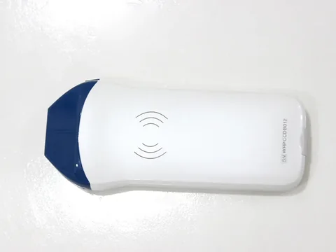
PICC/CVC workhorse · 5P
For intravenous therapy teams and PICC clinics doing mainly midlines and central venous access, plus difficult cannulation. Representative model 5P high-frequency linear wireless probe: 10.0–14.0 MHz, four depth settings of 20 / 30 / 40 / 55 mm, in-plane and out-of-plane guidance modes. Its typical uses read verbatim "nursing / access: PICC, CVC and other venous access; guidance for cannula placement, venepuncture for infusion and local drug administration".
Trade-off: 10.0–14.0 MHz is sufficient at this depth band, but the puncture assistance stops at "two guidance modes", with no automatic vessel measurement and no needle-tip enhancement. It solves "seeing the vessel", not "telling you where the tip has reached".
See this model's specifications →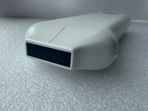
Difficult puncture · 6N with needle-tip cues
Where the vessels are fine, have been punctured many times, or sit deeper, and the tip needs to stand out on screen while the software reports calibre. Representative model 6N high-specification linear probe: 10.0–13.2 MHz, depth adjustable over 20–60 mm, all three puncture assistance items — in-plane guide line, out-of-plane guidance with automatic vessel measurement, needle-tip echo enhancement; 3 hours of battery life with wireless charging at any time.
Trade-off: what this tier adds is not frequency but cues. Needle-tip enhancement and automatic vessel measurement matter more to a less experienced operator; the cost is 200 g, somewhat heavier than the 5P.
See this model's specifications →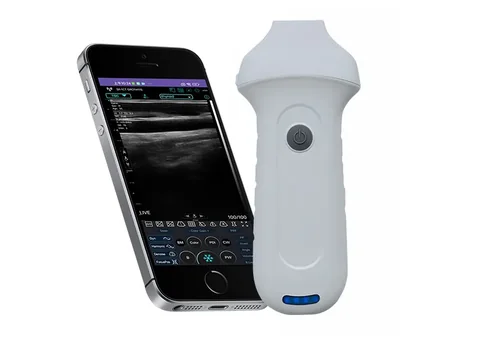
Very superficial and fine vessels · 7H
Paediatrics, neonates, the fine veins on the back of the hand and foot, small superficial veins — targets a few millimetres under the skin. Representative model 7H high-specification linear wireless probe: 16.0–20.0 MHz, depth adjustable over 10–40 mm, 180 g, in-plane guide line plus out-of-plane guidance with automatic vessel measurement.
Trade-off: 40 mm is a hard ceiling, and deep veins are simply out of reach. Its place in nursing work is not "the more advanced one" but a supplement dedicated to the very superficial category.
See this model's specifications →If a ward does not do many punctures and mostly needs cannula placement and infusion guidance, the cheapest configuration is none of the three above but the 5L or the 6L: 7.5–10.0 MHz, depths of 20 / 30 / 40 / 55 mm (the 6L adds a 100 mm setting), with the product page wording "venous access, cannula placement and local drug administration guidance, and other superficial procedures". Neither product page lists PICC or CVC among the uses, so match the selection to the procedures you actually perform.
One further point stands on its own: removal assessment uses a different category of device, not a probe. The BProbe-2 is 2.6 MHz with four-dimensional scanning and the BProbe-5 is 5 MHz with four-dimensional scanning, with the stated use "reference for the timing of catheter removal in postoperative patients with an indwelling urinary catheter, rapid bedside measurement". It measures urine volume in the bladder and has nothing to do with vessels, yet its place in the nursing workflow is the same as the three probes above — have a look before making the decision.
Five things that are easily overlooked
- Not every infusion needs ultrasound guidance. For a patient whose vessels are clear, palpable and cannulated first time, adding a device only adds a step. The cases that genuinely need guidance are a few specific groups: vessels that can neither be felt nor seen, a history of repeated failed punctures, the need for long-term placement, and strongly irritant drugs. Getting the indication clear matters more than studying the probe specifications in detail.
- The probe needs a sterile barrier, and our parameter tables do not say how. The parameter tables of the 12 products in this group list no sterile cover specifications, disinfection methods or ingress protection ratings. Access placement is an invasive procedure, so the barrier scheme for the probe and cable must follow the instructions for use and the hospital infection control protocols, not be applied from experience.
- In-plane and out-of-plane have to be practised, not just switched. In-plane shows the whole needle with the needle parallel to the probe's long axis; out-of-plane shows a cross-section of the tip with the needle perpendicular to the probe. The spatial sense of the two techniques is entirely different, and one person is often fluent in only one. Count training into the purchase, not just hardware.
- Screen placement decides technique. Nursing puncture is often done standing in the treatment room or at the bedside: put the phone on a table and you have to look down, put it on a stand and you need a free hand. In real use, screen height and angle affect the procedure more than 1 MHz of extra probe frequency.
- Battery life has to be counted per insertion. A PICC insertion, from assessment to dressing, commonly takes twenty or thirty minutes. The 5P is rated at about 3 hours (continuous-scanning basis) and the 7H at 1.5 hours. If four or five cases are booked in a day, the 7 series tier needs a charging dock, or choose the replaceable-battery 9L (two batteries at 1.5 hours each).
Four lines that cannot be crossed
This section matters more than every specification above.
- The device gives imaging reference; it does not promise a puncture outcome. Seeing a vessel's position, course and depth on screen does not mean the needle will go in. Success depends on vessel condition, patient cooperation, operator experience and other factors. We make no assurance about any puncture outcome, and neither the product pages nor this text contains any such statement.
- The qualifications and training requirements for access placement are outside the scope of the equipment. PICC and CVC are invasive procedures, and who may perform them and what training and authorisation are required is set by health administration and hospital policy. The device is responsible only for showing the vessel.
- Not every model lists PICC/CVC. The models whose product pages state "PICC, CVC and other venous access" explicitly among the applications are the 5P, 6L and 6N; the wording for the 5L is "venous access, cannula placement and local drug administration guidance, and other superficial procedures", with no mention of PICC or CVC. When selecting by use, check the product page wording item by item; do not reason that "they are all linear so they are all the same".
- Check the registration certificate's scope model by model. Which regions and which uses a given probe is registered for is governed by what the certificate states. Access placement involves intravascular procedures, so obtain the original certificate before purchase and check the "intended use" column — do not substitute the "applicable departments" line from a product page.
One more point: this page discusses the selection of ultrasound imaging equipment for nursing and vascular access settings. Nursing work also involves port maintenance, recognition and management of catheter-related complications, infection control and other steps; those are outside the scope of this page and should not be mixed into the same comparison table as the ultrasound configuration.
Common questions
Q: A nurse has never studied ultrasound — can they pick this up?
A: What a nurse learns for ultrasound-guided puncture is not ultrasound diagnosis but finding the vessel and recognising the tip: transverse scanning to find the vessel, longitudinal to see its course, the in-plane and out-of-plane techniques, and how to confirm the tip. Far less has to be mastered than for a physician performing an examination. The difficulty lies in technique and spatial sense, not in interpreting the image. Training is still essential, and no device shortens that step.
Q: Does venepuncture for infusion really need ultrasound?
A: Not all of it does. For a patient whose vessels are clear, palpable and cannulated first time, conventional technique is enough. The ones that need guidance are specific groups: not palpable, repeated past failure, long-term placement required, strongly irritant drugs. Concentrate those cases and the utilisation rate rises; use it on every patient in the ward and most of the time it is superfluous.
Q: How do I actually choose between in-plane and out-of-plane?
A: By what you need to confirm. In-plane shows the whole needle and is suited to judging whether it has wandered or transfixed the posterior wall; out-of-plane shows a cross-section of the tip and is suited to confirming the tip has entered the lumen. In practice you switch between them, so at selection confirm the probe has both modes — the parameter tables of the 5P and 6L both state "in-plane and out-of-plane modes" or "supporting in-plane and out-of-plane guide lines".
Q: Can PICC and cannula placement use the same probe?
A: Yes, provided the depth is covered. A cannula target sits 5–20 mm below the skin and PICC needs to see venous course at 30–50 mm, so a range like 20 / 30 / 40 / 55 mm covers it — both the 5P and the 5L have those settings. The difference between them is frequency: the 5P is 10.0–14.0 MHz and leans towards superficial detail, the 5L is 7.5–10.0 MHz and aims slightly deeper.
Q: Why does removal assessment count as a nursing scenario?
A: Because nursing carries out the removal, and "when to remove" needs a visible basis. Remove a urinary catheter too early and it has to be reinserted; too late and the indwelling time lengthens. The product pages of the BProbe-2 and BProbe-5 state exactly this: "for patients with an indwelling urinary catheter after surgery, to determine the timing of catheter removal". It is not the same category of device as a vascular probe, but it occupies the same place in the workflow: have a look before deciding.
Related reading
Vascular access and dialysis: choosing before, following up after
Also about vessels, but that page answers "is the access still flowing", which needs one depth tier deeper.
See the scenario →Intervention: how the needle is seen, and how to choose among four routes
The limits of needle-tip enhancement and magnetic navigation are set out in more detail on that page.
See the scenario →BProbe-2 wireless bladder volume measurement device: 2.6 MHz, four-dimensional
The device used for removal assessment; its product page lists uses across nine departments.
See the specifications →No dedicated sonographer: how a primary-care facility gets the most from portable equipment
Who operates it and how they are trained — nurse-led puncture guidance follows the same logic.
Read →List the punctures you perform and the depth tier settles itself
Tell us three things: which access types you mainly place, how many cases a day, and who operates. From those we can set the frequency band, the depth tier and whether needle-tip cues are needed; you can also download the selection table and compare for yourself first.
Download the selection table See other scenarios →The specifications described here are taken from the parameter tables published on this site. Their scope is reference information based on technical specifications; the content stated in the product registration certificate prevails. For contraindications and precautions, refer to the instructions for use. The product must be operated by a licensed physician with the appropriate qualifications.

