
Paediatric ultrasound: the difficulty is not organ size, it is whether the probe will sit flat
Press an adult abdominal probe onto a neonate's abdomen and the face is wider than the whole abdominal wall: the ends lift, gel cannot fill the gap, and the middle of the image is the part that goes blurred. Go the other way and put a high-frequency linear probe on the abdomen, and beyond twenty or thirty millimetres nothing comes back at all. What paediatrics needs is not a smaller machine but a probe that will sit flat on a small body — and one family of probes in the range on this site is built on exactly that idea: two active faces on one probe, microconvex for structures under a small contact area, linear for the superficial layer. This page sets out that division of labour, and marks separately the claims that cannot be made and the work this equipment cannot do.
Four constraints to work through first
The order matters: confirm the probe will sit flat before discussing how well it images.
The contact face is wider than the organ
The anteroposterior diameter of a neonate's or infant's abdomen is limited, so a standard convex face placed on it lifts at both ends, trapping air between probe and skin, and ultrasound does not cross air. The curved microconvex face is smaller, and this is stated plainly on the 5MN and 5ML product pages: a small microconvex contact area that sits well on a small body.
The range of depths is wide
Transcranial views of the brain have their target several centimetres down; subcutaneous tissue and superficial nodes sit within 20 mm. The two make opposite demands on frequency: depth needs low-frequency penetration, the superficial layer needs high-frequency resolution. That is why two faces on one probe comes closer to how paediatrics is actually practised than two separate probes.
The examining window is short
A child will cooperate for only so long, so switching on, connecting and adjusting the image all have to be quick. The wireless probes carry their own WiFi for image transmission and do not depend on the setting having a network; across the series the battery lasts about 3 hours of continuous scanning in normal working mode, and charging takes about 2–3 hours. Those two figures decide how many examinations can be done on one ward round.
One hand has to be left for the child
Holding position, soothing and shielding often take a hand. With the probe itself being the whole device and the screen a phone or tablet, that arrangement is more realistic at the bedside than wheeling a machine over. It connects to phones, tablets and Windows computers; Android devices can mirror to several screens over WiFi-Direct, while Apple devices are not currently supported.
Which probe for each of the routine paediatric examinations
The frequencies and depths below come from the specification tables for the 5MN and 5ML on this site and can be checked item by item.
| Examination | Probe type | Frequency · penetration depth |
|---|---|---|
| Brain (transcranial) | Microconvex | 5.0–7.0 MHz 60 / 90 / 120 / 160 mm |
| Abdominal organs | Microconvex | 5.0–7.0 MHz 60 / 90 / 120 / 160 mm |
| Lungs and pleura | Microconvex | 5.0–7.0 MHz from 60 / 90 mm |
| Paediatric hip screening | Microconvex | 5.0–7.0 MHz 60 / 90 mm |
| Superficial nodes, subcutaneous tissue | Linear | 10.0–12.0 MHz (5MN) 20 / 40 / 60 / 80 mm |
| Musculoskeletal, nerves (cooperative older children) | Linear | 10.0–12.0 MHz / 7.5–10.0 MHz (5ML) 20 / 40 / 60 / 80 mm |
| Vessel localisation and puncture guidance | Linear + guide line | 10.0–12.0 MHz in-plane and out-of-plane modes |
The first four rows all use the same face, and that is no accident. The microconvex depth settings run from 60 mm to 160 mm, which covers the transcranial and abdominal depths of neonates through to preschool children; beyond that, past 200 mm, is work for an adult convex probe, which paediatrics does not need and should not use — drop the frequency and the superficial resolution goes with it.
The last three rows use the other face. The linear range at 10.0–12.0 MHz with 20–80 mm depth covers subcutaneous tissue, superficial nodes, and musculoskeletal and nerve work in older children who can cooperate. Switching is by button on the same probe, with nothing to change and no second machine. This is also the substantive difference between the 5MN and the 5ML: the 5MN's linear face is 10.0–12.0 MHz, leaning superficial; the 5ML's is 7.5–10.0 MHz, aimed a little deeper. The microconvex section and the depth settings are identical on both, and the selection note in our catalogue says exactly that — the difference from the 5ML is in the linear frequency band.
Three common configurations, chosen by who is using it
The three tiers correspond to three settings; they are not a cheaper-to-better ladder.
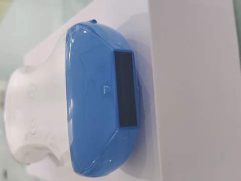
Superficial-led · One 5MN
For neonatology and paediatric clinics whose work is mainly brain, abdomen and lungs, with some superficial node and subcutaneous work. Representative model: the 5MN microconvex plus linear dual-head wireless probe — microconvex 5.0–7.0 MHz, depth 60 / 90 / 120 / 160 mm; linear 10.0–12.0 MHz, depth 20 / 40 / 60 / 80 mm; all modes (B, B/M, Color, PDI, PW) and both in-plane and out-of-plane guide lines.
The trade-off: the high-frequency linear face gives more superficial detail at the cost of seeing nothing beyond 80 mm. Deeper structures are handled by the microconvex half, with a button to switch — which is the entire reason the arrangement exists.
See the specifications →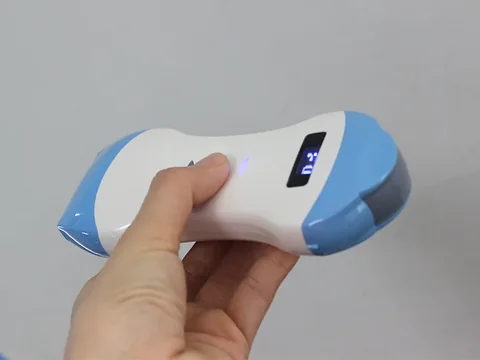
A little deeper · One 5ML
Same brain and abdomen work, but where the superficial section needs to reach slightly deeper structures, or the operator is more used to the feel of the 7.5–10.0 MHz band. Representative model: the 5ML microconvex plus linear dual-head wireless probe — the microconvex side is identical to the 5MN (5.0–7.0 MHz, four settings from 60 to 160 mm), while the linear face drops to 7.5–10.0 MHz at the same 20 / 40 / 60 / 80 mm depths.
The trade-off: a lower linear frequency gives less detail in the very top layer than the 5MN, but penetrates the 40–80 mm band more comfortably. Neither is better than the other; they lean different ways.
See the specifications →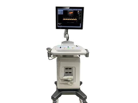
A fixed installation · Console with a microconvex probe
Where the department has a fixed examining room, staff work in shifts and daily volume is steady, a console comes into consideration. Taking the C300 trolley system as an example: the standard configuration includes an abdominal convex probe and a built-in battery, the maximum scanning depth is 24 cm, and the options list explicitly includes a linear probe, an endocavity probe, a microconvex probe, a phased-array probe and a needle guide.
The trade-off: a console gives a full screen, measurement software and a reporting chain, but it takes floor space and needs power; and the microconvex probe is an option, so it has to appear as its own line on the configuration list — otherwise only the standard probe arrives.
See this configuration →There is a lighter route as well: if the need is a quick look at the bedside and a console already handles formal examinations, a single-face microconvex probe is enough and the dual head is unnecessary. The test is simple — list the department's paediatric examinations over the past month in two columns, depth and contact area. If the great majority fall within 160 mm and superficial work is under a fifth, the second face is sitting idle.
Conversely, if superficial nodes, subcutaneous tissue and musculoskeletal work make up more than half, the dual head earns its place: what it saves is not the cost of a probe but the changing of parts and re-focusing at every examination.
Four things that often get missed
- Gel temperature matters as much as anything else. Neonates lose heat quickly from the skin, and once an examination runs long the child cries, position shifts, and the image shifts with it. Warming the gel and splitting the examination into stages cost nothing, and affect the image as much as changing probe does.
- Screen size decides who can actually see. The wireless image appears on a phone or tablet — small, not very bright, and prone to reflections in a well-lit ward. If judgements are really to be made at the bedside, the size of the tablet and shading it are worth solving before any probe specification.
- Mirroring depends on the device. Android devices can mirror to several screens over WiFi-Direct; Apple devices are not currently supported. If mirroring is needed for teaching, consultation or talking with a family, confirm in advance which kind of device is being used rather than discovering on the spot that nothing can be cast.
- Battery life is quoted for continuous scanning. The specification says about 3 hours of uninterrupted scanning in normal working mode. A real ward round is intermittent, and standby draws power too, so for a whole morning a power bank or a wireless charging dock goes in the bag as well.
Four lines not to cross
This section matters more than every specification above it.
- Paediatric hip screening is an application, not a technique. The product pages on this site list it among the typical uses in neonatology and paediatrics, but how screening is performed, which view is used and which angles are measured belong to clinical guidance and departmental protocol, not to equipment selection. What the equipment contributes is the 5.0–7.0 MHz band and 60–160 mm depth.
- The scope of the registration certificate has to be checked device by device. For any given probe, which body regions and which patient groups the certificate covers is determined by what the certificate says. Paediatric use involves age and body size, so the certificate itself should be pulled and its scope read before purchase — a product page's list of subjects is not a substitute.
- One scan does not close a diagnosis. Imaging findings in the brain, lungs and hip all have to be judged together with history, signs and other investigations. The device supplies the image; the judgement belongs to the physician.
- Paediatrics is not an adult protocol scaled down. Shrink an adult abdominal protocol proportionally onto a child and what you get is a combination of too low a frequency and too large a contact face. Configuring by body size means going back to two things — contact face and depth range — rather than guessing from age or weight.
One further point: this page is about selecting ultrasound imaging equipment. Paediatric practice also uses monitoring and therapeutic equipment of other kinds; those are outside its scope and should not be mixed into the same comparison table as ultrasound configuration.
Common questions
Q: What makes paediatric ultrasound harder than adult?
A: Two places, and neither is that the organs are small. The first is the contact face — a neonate's anteroposterior diameter is limited, so an adult convex face lifts at both ends and gel will not fill the middle. The second is the depth range — the brain has to be seen several centimetres down, while subcutaneous tissue and superficial nodes sit within 20 mm, one needing low-frequency penetration and the other high-frequency resolution. The microconvex-plus-linear dual head is built around exactly those two constraints.
Q: What exactly is the difference between the 5MN and the 5ML?
A: Only the linear frequency band. The 5MN linear face is 10.0–12.0 MHz, leaning superficial; the 5ML is 7.5–10.0 MHz, aimed a little deeper. Both have a 5.0–7.0 MHz microconvex face with depths of 60 / 90 / 120 / 160 mm and linear depths of 20 / 40 / 60 / 80 mm, and are otherwise the same. Which one to choose depends on whether your superficial work needs finer detail or a little more depth.
Q: For brain and abdomen only, is a dual head needed?
A: Not necessarily. Where superficial work is a small share, a single-face microconvex probe is enough and the budget is better spent on screen and battery. The dual head's value is that nothing has to be changed on the same device, and that value rises with the share of superficial work.
Q: With a console already in the department, is a wireless probe still needed?
A: It depends on where it will be used. A console is efficient for formal examinations in the examining room, but moving it to a ward or a bedside is limited by power and by corridors. A wireless probe is the whole device, connecting to a phone or tablet for the image, and what it adds is the look at the bedside. The two do not replace each other.
Q: Can a console be supplied with a microconvex probe?
A: Yes, but it has to be written into the configuration list. On the C300, the options explicitly include a microconvex probe; the C5T options list one too. The standard configuration usually includes only an abdominal convex probe, so the microconvex is a separate item, and leaving it off the order means a separate order later.
Related reading
Musculoskeletal ultrasound: a trade-off table for frequency and depth
Superficial work in paediatrics follows the same logic, one depth band shallower.
See the scenario →Primary care screening: working back from a day's caseload
If paediatrics is only one item in the clinic, start with the approach on this page.
See the scenario →Handheld, portable or trolley ultrasound: start with where it will sit
Form factor before specifications — where bedside and examining room divide for paediatrics is set out here.
Read →Using portable ultrasound well without a dedicated sonographer
Who operates, how they are trained, when referral is mandatory.
Read →Sort the examinations by depth and the probe answers itself
The Selection Guide section of this site carries a comparison table arranged by examination site and penetration depth, for discussion within the department; or simply tell us the tasks and we can suggest a configuration against the actual caseload.
Download the selection guide See other scenarios →The specifications in this article are taken from the product specification tables on this site. This document is a reference explanation based on technical parameters; where it differs, the content stated on the product registration certificate takes precedence. For contraindications and precautions, see the product instructions for use. The product must be operated by a licensed physician with the appropriate qualifications.

