
Skin and subcutaneous tissue: in this tier, choosing a probe means choosing how deep you can see
On the musculoskeletal, abdominal and cardiac pages, selection balances frequency, depth, element count and assistance functions. The skin tier is far simpler and far harsher: the higher the frequency, the shallower the penetration — there is no exception and no parameter tweak that gets around it. The 18–24 MHz probe stops at 25 mm, not one millimetre further; the 10–13.2 MHz probe reaches 60 mm, at the cost of the most superficial detail. So this page does not discuss "which is better", only "at how many millimetres below the skin is the layer you need". Our line has 7 probes covering skin and superficial work, in four depth bands.
Four hard constraints in skin ultrasound
The first is a law of physics; the other three come from the bedside.
Frequency buys depth, one for one
The 8S is 18.0–24.0 MHz with only four depth settings — 10 / 15 / 20 / 25 mm; the 8H is 16.0–20.0 MHz at 10 / 20 / 30 / 40 mm; the 8N is 10.0–13.2 MHz at 20 / 30 / 40 / 60 mm. Three models, nine numbers, and the whole trade-off of this tier is stated.
The shallower it is, the harder it is to couple
Looking at structures within 10 mm, there must be no air at all between probe and skin, and the probe must not press hard — press down and the target is flattened and displaced. A high-frequency probe has a narrow acoustic window and loses the image at the slightest tilt. This tier is far more sensitive to gel volume and hand pressure than the abdomen.
An uneven surface cannot be imaged
Nodules, ulcers, scars, dressings and hair all leave gaps between probe and skin. High-frequency sound is totally reflected at an air interface, and the image goes black. With such patients you either fill with a generous layer of gel or add a water bag or ultrasound pad — a matter of technique, not something the device can solve.
Superficial examinations are short
A skin examination usually takes a few minutes, and a clinic may see many patients in a day. So this group is positioned as "pick it up and go": the 8 series weighs 180 g, the 9 series 230 g, and the bodies are 140 or 156 mm long, so both fit in one hand. On battery life, the 8 series has an internal battery good for 1.5 hours (3 hours with the extended version), and the 9 series two batteries at 1.5 hours each.
Four depth bands, four tissue layers
Frequencies and depths below are taken from our published parameter tables and can be checked item by item against the model.
| Depth band | Model · frequency · depth settings | Which layer it reaches |
|---|---|---|
| 10–25 mm |
8S / 9S
18.0–24.0 MHz 10 / 15 / 20 / 25 mm |
Epidermis, dermis, superficial subcutaneous tissue |
| 10–40 mm |
8H / 9H
16.0–20.0 MHz 10 / 20 / 30 / 40 mm |
Full thickness of skin plus subcutaneous fat |
| 20–60 mm |
8N / 9N
10.0–13.2 MHz 20 / 30 / 40 / 60 mm |
Deep subcutaneous tissue, fascia, larger areas |
| 20–60 mm, wide field |
5W
7.5–10.0 MHz 20 / 40 / 60 mm |
Overall survey of a large area |
The 25 mm in the top row is a hard boundary. In the 18.0–24.0 MHz tier the depth settings end at 25 mm — not "you can set it deeper but the image degrades", but simply that there is no deeper setting. If your patient has a thick subcutaneous fat layer, or the target sits around 30 mm, this probe is unsuitable on principle.
Conversely, the 5W in the bottom row is the only one in this group that is not ultra-high frequency: 7.5–10.0 MHz, but with 256 elements and a body width of 80 mm (other models are 55 or 60 mm), giving a wide acoustic window. Its value is not frequency but how much it covers in one view — for extensive subcutaneous oedema, skin graft areas or widespread subcutaneous emphysema, where an overall judgement is needed, a wide window is more useful than high frequency.
Why there are two nearly identical series
This is the part of our line that most often leaves people stuck.
| Item | 8 series | 9 series |
|---|---|---|
| Frequency and depth | Identical (8N to 9N, 8H to 9H, 8S to 9S, item by item) | |
| Elements and channels | 192 elements, 64 channels, the same in both series | |
| Weight | 180 g | 230 g |
| Battery | Internal, 1.5 hours in the standard version 3 hours with the extended battery version |
2 batteries supplied, replaceable 1.5 hours each |
| Body length | 140 mm | 156 mm |
| Puncture assistance | In-plane guide line plus out-of-plane guidance and automatic vessel measurement | In-plane guidance plus out-of-plane guide line |
This table says three things. First, the difference between 8 and 9 is not in the image: frequency, depth, elements and channels are all identical, so on paper they ought to be "the same specifications, therefore the same thing". Second, the difference is in how they are powered: the 8 series has one internal battery, and for longer running you choose the extended battery version; the 9 series carries two detachable batteries, so you swap one out and carry on without waiting to recharge. Third, weight and length are the price: the extra structure for a replaceable battery makes the 9 series 50 g heavier and 16 mm longer.
So which series to choose has nothing to do with image clarity and everything to do with how a day runs. A clinic seeing a dozen or so patients with time to recharge in between is better served by the lighter, better-balanced 8 series; a day of back-to-back work, or carrying it to the ward, makes the 9 series' swap-without-stopping more practical.
One detail also deserves attention: the 9 series actually has one fewer puncture assistance item than the 8 series. The 8 series parameter table reads "in-plane puncture guide line, out-of-plane puncture guidance and automatic vessel measurement"; the 9 series reads "in-plane puncture guide function, out-of-plane puncture guide line function", with no automatic vessel measurement. That runs against the intuition that 9 outranks 8, so check the parameter table wording item by item rather than reasoning from the model number.
Three set-ups, chosen by how deep you need to see
Settle the depth first, then choose 8 or 9 within that band.
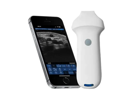
The skin itself · 8S, the shallowest tier
Where the target is the skin and the few millimetres immediately beneath it, and layer boundaries have to be resolved. Representative model 8S ultra-high-frequency linear probe: 18.0–24.0 MHz, four depth settings of 10 / 15 / 20 / 25 mm, 192 elements and 64 channels, 180 g. The applications on its product page read verbatim "observation of skin, subcutaneous tissue and other superficial structures".
Trade-off: 25 mm is an absolute ceiling. Within this layer it gives the highest resolution, but it cannot see the deeper part of subcutaneous fat, nor the fascia. Using it on a patient with a thick fat layer is like reading a distant view through a magnifying glass.
See this model's specifications →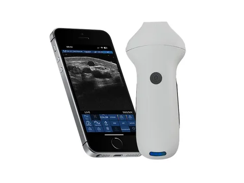
Skin plus subcutaneous · 8H, the balanced tier
Where you need the skin layers and also the subcutaneous fat over a wider field, without changing probes to get it. Representative model 8H ultra-high-frequency linear wireless probe: 16.0–20.0 MHz, four depth settings of 10 / 20 / 30 / 40 mm, 192 elements and 64 channels, 180 g, with out-of-plane guidance and automatic vessel measurement among the assistance functions.
Trade-off: slightly less of the most superficial detail than the 8S, in exchange for 15 mm of extra depth. This is the most general choice in the category — a clinic whose patients vary widely in build and whose examinations vary widely in type usually takes this one.
See this model's specifications →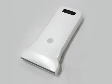
Large-area survey · 5W, wide acoustic window
Where the object is a whole subcutaneous region and a wider field has to be covered in one view. Representative model 5W ultra-wide linear wireless probe: 7.5–10.0 MHz, depths of 20 / 40 / 60 mm, 256 elements, body width 80 mm (other models 55–60 mm), 250 g, 2.5 hours of battery life, with needle-tip echo enhancement among the assistance functions.
Trade-off: frequency drops to 7.5–10.0 MHz and the most superficial detail is the weakest in this group; what you get is width and element count. It is not for close reading of skin layers but for the overall question of what is going on across a region.
See this model's specifications →One band does not get its own card: the 8N and 9N, at 10.0–13.2 MHz with depths of 20 / 30 / 40 / 60 mm, pushing the ceiling to 60 mm. If your patients are generally obese and the target usually sits around 40 mm below the skin, this is the right band — the lowest frequency and deepest penetration in the group, with superficial detail inevitably sacrificed.
Five things that are easily overlooked
- High frequency is more sensitive to attenuation. At the same frequency, oedematous, scarred and fibrotic tissue attenuates the sound faster, and the lower half of the image darkens with blurred boundaries. The remedy is to drop one frequency tier (from 18–24 MHz to 16–20 MHz, for example), not to turn up the gain — raise the gain and the noise comes up with it.
- The harder you press, the shallower the target looks. Superficial structures are thin to begin with, and pressing the probe thins the subcutaneous fat and pushes or flattens the target, so the measured thickness is wrong. The correct approach is to touch without pressing, filling with plenty of gel and letting the probe float on the skin.
- Hair and dressings have to be dealt with. Over body hair, dressings or ointment, sound is totally reflected at the air interface. Shaving, removing the dressing or adding an ultrasound pad is routine preparation for this kind of examination; skip it and the screen is black.
- On the 8 series you need the extended battery version to get 3 hours. The parameter table reads "1.5 hours in the standard version, 3 hours with the extended battery version". If the purchase order names the model but not the battery version, what arrives is the 1.5 hour one. The 9 series is "2 batteries supplied, 1.5 hours each", and "2 batteries" has to be written in explicitly.
- The price of a wide window is the contact surface. The 5W body is 80 mm wide, so on small areas such as fingers, the dorsum of the foot or the face it overhangs and lifts at both ends. A wide field is valuable on flat, large areas, not on irregular ones.
Four lines that cannot be crossed
This section matters more than every specification above.
- "Observation" is not "characterisation". The applications wording for all 7 products in this group is the same single sentence — "observation of skin, subcutaneous tissue and other superficial structures". Observation means seeing layers, thickness, echogenicity and boundaries; what those findings mean and whether anything should be done is a clinical judgement. The device does not carry that layer, and this text contains no statement about the nature of any lesion.
- Frequency and depth are physical constraints, not configuration options. 18.0–24.0 MHz reaches 25 mm and no further; there is no "adjusting the depth" to see 40 mm. People ask whether the limit can be released in software — it cannot: what appears on screen afterwards is noise, not an image. To see deeper you have to change to a lower-frequency probe.
- Probe contact surface and skin condition affect each other. Over ulcers, broken skin, infection or dressings the probe must not be placed directly on the surface, and sterile barrier and protection measures follow hospital protocol. Our parameter tables do not list disinfection methods or ingress protection ratings for each model; that has to be checked in the instructions for use.
- Check the registration certificate's scope model by model. Which regions and which uses a given probe is registered for is governed by what the certificate states. Skin and superficial work involves the body surface, so obtain the original certificate before purchase and check the "intended use" column — do not substitute the "applicable departments" line from a product page.
One more point: this page discusses the selection of ultrasound imaging equipment for skin and subcutaneous settings. Dermatology also involves dermoscopy, confocal microscopy and other imaging methods; those are outside the scope of this page and should not be mixed into the same comparison table as the ultrasound configuration.
Common questions
Q: How deep can skin ultrasound see?
A: It depends which probe. The shallowest in this group is the 8S at 18.0–24.0 MHz with depth settings of 10 / 15 / 20 / 25 mm; the most general is the 8H at 16.0–20.0 MHz with 10 / 20 / 30 / 40 mm; to reach 60 mm you need the 8N at 10.0–13.2 MHz with 20 / 30 / 40 / 60 mm. The three are three different depth lines, not three grades of precision.
Q: The 18–24 MHz tier has the highest resolution — why not just buy that one?
A: Because it stops at 25 mm. Higher frequency means shallower penetration; that is the physics of ultrasound, not a product defect. With a thick subcutaneous fat layer, or a target around 30 mm, this probe cannot see it on principle — not "the image is a bit worse" but "that thing is not on the screen". Settle the depth requirement first, then discuss frequency.
Q: In oedematous or scarred tissue, why is the lower half of the image so dark?
A: Sound attenuates as it travels through tissue, and oedematous, fibrotic and scarred tissue attenuates faster than normal subcutaneous fat, so the signal returning from deeper is weaker. The remedy is to drop one frequency tier (from 18–24 MHz to 16–20 MHz, for example), or to use the wide-window 5W for an overall judgement first and then switch to high frequency for detail. Raising the gain does not solve it; it amplifies the noise as well.
Q: What is the 5W's wide field actually for?
A: For large areas with a flat surface. Its body is 80 mm wide (other models 55–60 mm) with 256 elements, covering a wider strip in one view, which suits an overall survey of a subcutaneous region — the distribution of extensive subcutaneous oedema, a skin graft area, subcutaneous emphysema. Narrow sites such as fingers, the dorsum of the foot and the face do not suit it; the probe lifts at both ends.
Q: How does this page divide work with the musculoskeletal page?
A: The musculoskeletal page is concerned with tendons, ligaments, joint spaces and effusions, at depths from 10 mm to 60 mm, and divides selection into three tiers by how deep the structure lies; this page is concerned with the layers of skin and subcutaneous tissue themselves, with depth concentrated within 25 mm. The models overlap (the 8H appears on both), but the questions differ: one is "how is this tendon", the other is "how thick is this layer of skin".
Related reading
Musculoskeletal: narrowing the field to three models by depth
The same "frequency buys depth" logic, applied to tendons, ligaments and joints.
See the scenario →Paediatrics: small contact surfaces and a wide spread of layers
Children's skin and subcutaneous tissue are thinner, and the superficial tier is used differently in paediatrics.
See the scenario →8S ultra-high-frequency linear probe: 18.0–24.0 MHz
The shallowest probe in this tier; its product page gives the full four depth settings from 10–25 mm.
See the specifications →5W ultra-wide linear wireless probe: 256 elements
The one with both a wide acoustic window and needle-tip enhancement; specifications and usage on that page.
See the specifications →Measure one thing first: at how many millimetres sits the layer you need
Once you have that number, the frequency and depth tier follow. If you are unsure, tell us the body regions and approximate depths you see most often and we can base configuration advice on that; you can also download the selection table and compare for yourself first.
Download the selection table See other scenarios →The specifications described here are taken from the parameter tables published on this site. Their scope is reference information based on technical specifications; the content stated in the product registration certificate prevails. For contraindications and precautions, refer to the instructions for use. The product must be operated by a licensed physician with the appropriate qualifications.

