
Ophthalmic ultrasound: A-scan and B-scan are two jobs, not two modes of one machine
Ophthalmic ultrasound differs from other specialties in one respect: its main job is not looking but measuring. The eye is a small organ a few centimetres across, and a general-purpose probe placed against it is all near-field blind zone, which is why a dedicated small probe is needed. And the A-scan and B-scan on such a device do two quite different jobs — the B-scan shows the vitreous, retina and other posterior segment structures; the A-scan measures axial length, anterior chamber depth and lens thickness, and the numbers it produces directly determine the intraocular lens power. Discuss the two as one thing and the configuration will certainly be wrong.
Four constraints to work through first
Decide first whether the job is viewing the posterior segment or taking measurements; the rest becomes much clearer.
The probe has to be an ophthalmic one
The eye is small and superficial. Put a general abdominal or high-frequency linear probe against it and the near-field blind zone covers exactly the region you need to see. The dedicated small ophthalmic probe (10 MHz on the models on this site) is built for that distance.
The numbers go into a formula
Axial length, anterior chamber depth and lens thickness are not reference values; they are inputs to the intraocular lens power calculation. So the first requirement of this equipment is that it measures accurately and reproducibly.
The same eye has to be comparable
Biometry results have to be comparable across repeat visits on the same eye. Method matters as much as the device: the same position, the same fixation, the same measurement conditions.
Corneal thickness is another item
Preoperative assessment and postoperative evaluation in refractive surgery look at central and peripheral corneal thickness, and the models on this site measure to ±0.05 mm. This and axial length measurement are often done on the same dedicated device.
Which category each of the five routine clinic examinations falls into
The figures below come from the technical descriptions of the ophthalmic products on this site and can be checked item by item against the product pages.
| What is viewed or measured | What is used | Key figures |
|---|---|---|
| Vitreous opacities, retinal detachment, orbital lesions (morphology) | B-scan | Three imaging modes: normal, vitreous enhanced, retinal |
| Anterior chamber depth, lens thickness, axial length | A-scan | 10 MHz small probe, penetration depth 40 mm |
| Intraocular lens power calculation | A-scan plus calculation formula | Measurement modes for phakic, aphakic and pseudophakic eyes |
| Central and peripheral corneal thickness | A-scan pachymetry | Measurement range 15–40 mm, precision ±0.05 mm |
| Macular localisation and fixation monitoring | A-scan (macular tracking) | Macular recognition, automatic central and peripheral compensation |
What matters most in this table is the difference between the first two rows. A B-scan gives an image — whether there are opacities in the vitreous, whether the retina is detached, whether there is an orbital mass: form and spatial relationships. An A-scan gives numbers — an echo sequence along the axis, from which anterior chamber depth, lens thickness and axial length are read, and those numbers go into the formula that gives the intraocular lens power. The first is a matter of looking; the second of algorithms and protocol.
The third row is the step many people miss when configuring equipment: A-scan measurement modes distinguish phakic, aphakic and pseudophakic eyes. For a patient who has had cataract surgery, choosing the wrong mode makes the reading unusable. This is not a bolded line in the specification table at purchase time, but it comes up daily in use.
The fourth row's precision deserves separate attention: ±0.05 mm, measurement range 15–40 mm. Preoperative and postoperative assessment in refractive surgery turns on differences of that order — which is why pachymetry and general abdominal ultrasound are not the same class of equipment at all.
Three common configurations, chosen by the examining room
The three differ mainly on whether a mainframe is built in and whether a dedicated measurement station is needed.
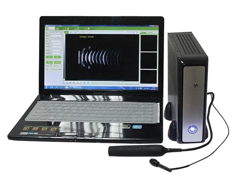
Added to a consulting room · No mainframe
For tight spaces and moving between consulting room and bedside. Representative model: the SAB-200 ophthalmic A/B scanner — a probe plus companion software, used by connecting to a computer and installing the software, with no separate mainframe.
The B-scan section offers three modes — normal, vitreous enhanced and retinal — for viewing the vitreous, retina and other intraocular structures; the A-scan section measures anterior chamber depth, lens thickness and axial length, with the results used for intraocular lens power calculation. Images can be stored, managed and edited in the software, and reports generated as needed.
See the specifications →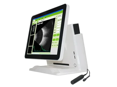
Dedicated examining room · Screen and battery built in
For a fixed examining room doing continuous examinations every day. Representative models: the SAB-500 and the SAB-600 — 15-inch colour LCD, imaging modes B, B+B, B+A and A, a built-in battery good for 4 hours, and a unit measuring 362 mm × 340 mm × 60 mm at 2.5 kg.
The A-scan uses an imported 10 MHz small probe that can be hand-held, immersed or slit-lamp mounted; scanning modes include contact and immersion, penetration depth is 40 mm, and measurement modes cover phakic, aphakic and pseudophakic eyes. Custom report templates are supported. The two units are close in overall specification but list different probe figures, so confirm against the actual configuration when choosing.
See the specifications →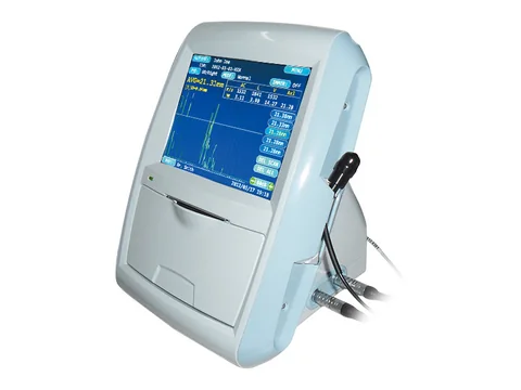
Dedicated measurement station · Corneal thickness
For institutions with high volumes of pre-cataract biometry, or that perform refractive surgery. Representative model: the SAP-100 ophthalmic A-scan pachymeter — 10 MHz A-scan probe with a built-in light source, measurement range 15–40 mm, precision ±0.05 mm, with macular tracking.
It measures anterior chamber depth, lens thickness, vitreous length and total length with their averages, applies automatic compensation for central and peripheral corneal thickness, and serves preoperative examination and postoperative evaluation in refractive surgery. This tier is the one that gets measurement right; it does not cover posterior segment morphology.
See the specifications →The three do not replace one another. Clinically the A-scan (measurement) and B-scan (morphology) are often needed together, which is why a combined A/B unit such as the SAB series is a common choice; institutions with very high measurement volumes add a separate pachymetry station alongside, so that preoperative measurement can be scheduled apart.
Four things that often get missed
- Measurement conditions have to be fixed. Biometry depends on position and on the patient's cooperation with fixation. For repeat measurements on the same eye, keep the conditions the same so the results are comparable — this directly affects follow-up decisions and is easier to overlook than any specification.
- Choose the wrong measurement mode and the number is worthless. Phakic, aphakic and pseudophakic are three different modes. After cataract or refractive surgery the mode has to change with the patient. This is where daily errors usually come from, not from the device.
- Contact and immersion each have their setting. Contact is quick but touches the cornea; immersion uses a coupling medium and needs cooperation, and holds a steadier image. Where one device supports both, write the choice into the operating protocol rather than leaving it to each operator.
- Settle the report template early. An ophthalmic report has to be readable directly by the surgeon, with consistent item order, units and reference notes. Where the device supports custom templates, configure them to departmental habit at installation rather than standardising six months later.
Three lines not to cross
This section matters more than every specification above it.
- Ultrasound does not replace fundus examination. Ophthalmoscopy, optical coherence tomography and fluorescein angiography provide information ultrasound cannot. Ultrasound's value is that it can see past a hazy ocular medium — vitreous haemorrhage, dense cataract — but it is not a substitute for examining the fundus.
- Measurements have to be read against the state of the eye. Axial length, anterior chamber depth and corneal thickness need cautious interpretation in eyes with previous surgery, silicone oil filling or corneal disease, and the choice of formula and mode has to be adjusted accordingly. The device produces the number; whether it can be used is the physician's judgement.
- Diagnosis of posterior segment disease rests with the ophthalmologist. Finding signs such as retinal detachment, vitreous opacity or an orbital mass is screening and flagging; the final diagnosis and management plan are determined by a qualified ophthalmologist. The device and the operator supply images and clues.
One further point: ophthalmic ultrasound probes are purpose-built. A general ultrasound system with a small probe swapped in will not do — the near-field behaviour, penetration depth (40 mm for the A-scan on the models here) and measurement modes are all designed around the scale of the eye. Data from a general-purpose device forced into ophthalmic measurement cannot go into intraocular lens calculation.
Common questions
Q: What actually is the difference between an A-scan and a B-scan?
A: An A-scan gives an echo sequence along one axis and outputs numbers — anterior chamber depth, lens thickness, axial length — which go into the formula for intraocular lens power. A B-scan gives a cross-sectional image showing the form and spatial relationships of the vitreous, retina and orbit. One device can carry both functions, but they answer different questions and do not replace each other.
Q: For pre-cataract measurement only, what is needed?
A: The A-scan section is the core. You need the 10 MHz small probe, 40 mm penetration depth, and support for phakic, aphakic and pseudophakic measurement modes. A probe-plus-software format such as the SAB-200 delivers measurement and reporting from a connected computer; if the posterior segment also has to be viewed, choose an all-in-one unit with a B-scan.
Q: Are the probes dedicated? Can a general ultrasound probe be used instead?
A: No. The eye is small and superficial, the near-field blind zone of a general probe covers exactly the target region, and neither the penetration depth nor the measurement modes are designed at that scale. Axial length measured on a general-purpose device cannot go into intraocular lens calculation, and that should be clear at the purchasing stage.
Q: Contact or immersion — how to choose?
A: It depends on working habits and how well the patient cooperates. Contact is quicker but needs topical anaesthesia and preparation of the cornea; immersion images through a coupling medium and requires the patient to hold fixation. The A-scan on the SAB-500 and SAB-600 here supports both scanning modes; which one is used should be written into the departmental protocol and kept consistent.
Q: What is corneal thickness measurement for?
A: Mainly preoperative assessment and postoperative evaluation in refractive surgery. Both central and peripheral thickness are needed, and precision at the ±0.05 mm level is what it takes. The SAP-100 provides automatic central and peripheral compensation, a measurement range of 15–40 mm, and macular tracking.
Related reading
Handheld, portable or trolley ultrasound: start with where it will sit
Ophthalmic equipment mostly lives in consulting and examining rooms, where form and placement matter just as much.
Read →Using portable ultrasound well without a dedicated sonographer
Ophthalmic ultrasound depends on protocol; this page is about workflow rather than specifications.
Read →Ophthalmic B-scanners: a model-by-model comparison
The detailed specifications of the SAB and SAP series can be compared directly in the specification tables.
See the product section →Primary care screening: working back from a day's caseload
If ophthalmology is only one item in the clinic, start with the approach on this page.
See the scenario →Decide first whether biometry is needed, then discuss models
A configuration for posterior segment imaging alone differs substantially from one that also has to produce preoperative measurement data. The Selection Guide section of this site carries a comparison table arranged by examination, for discussion within the department.
Download the selection guide See other scenarios →The specifications in this article are taken from the product specification tables on this site. This document is a reference explanation based on technical parameters; where it differs, the content stated on the product registration certificate takes precedence. For contraindications and precautions, see the product instructions for use. The product must be operated by a licensed physician with the appropriate qualifications.

