
Vascular access ultrasound: before the needle goes in you ask "which vessel", afterwards you ask "is it still flowing"
Access ultrasound looks like one thing but is really two. Before the procedure it has to answer "is this vessel large enough, how many millimetres below the skin is it, is there a companion structure alongside" — the answer decides where the needle goes. Afterwards it has to answer "is there still flow in this access, has the lumen narrowed, is there material on the wall" — the answer decides how much longer the access will last. The two questions call for different depths, which is why one department often keeps two probes rather than one. Our line has 7 probes covering vascular access and dialysis; this page arranges them by depth setting and marks separately the parts the parameter tables do not cover.
Four hard constraints in access ultrasound
The first two decide whether the image is usable; the last two decide whether the device really gets picked up every day.
Depth settings eliminate half the models first
Access vessels sit between a few millimetres and a few centimetres below the skin. That range looks shallow but spans three tiers: 0–40 mm, 0–60 mm and 0–100 mm. In this group on our site, the 7H has a depth of 10–40 mm, the 6N 20–60 mm, and the 7L and 9L 20–100 mm. The three tiers are not a difference in precision but in whether you can see at all.
Frequency and penetration pull against each other
The 7H is 16.0–20.0 MHz and shows the layers of the vessel wall in the finest detail, but beyond 40 mm it sees nothing; the 7L is 7.5–10.0 MHz and reaches 100 mm, at the cost of wall detail. The commonest mistake in access ultrasound is to take a high-frequency probe to a deep access and then conclude that "it cannot be seen" — the depth setting was insufficient, not the image quality poor.
Puncture assistance is not on every probe
Across these 7 probes the puncture assistance is worded in four ways: only "in-plane and out-of-plane modes" (5P); up to "automatic vessel measurement" (7H, 7L, 7N); plus a further "needle-tip echo enhancement" (6N); and with "magnetic navigation display of needle displacement and angle" (6X). These four are verifiable differences, not the same sentence reworded.
Dozens of times a day
Access assessment in a dialysis centre is highly repetitive work, and the device has to be picked up, put down and picked up again with one hand. Weights in this group: the 7 series at 180 g, the 6N and 6X at 200 g, the 9L at 230 g. On battery life, the 6N and 6X are rated at 3 hours, some 7 series models at 1.5 hours, and the 9L is two batteries at 1.5 hours each.
Three actions, and the depth each one needs
Frequencies and depths below are taken from our published parameter tables; the three columns correspond to three points in access management.
| What you are doing | What you need to see | Frequency · depth needed |
|---|---|---|
| Before: choosing the vessel | Calibre, depth below skin, course, presence of companion structures | 10.0–13.2 MHz or 7.5–10.0 MHz from 20–60 mm |
| During: catheter placement and puncture | Needle-tip position, shaft course, whether the lumen has been transfixed | 10.0–14.0 MHz or 7.5–10.0 MHz 20–55 mm settings |
| After: access follow-up | Lumen, flow, wall and surrounding tissue | 7.5–10.0 MHz 20–100 mm |
The third row is where this page divides from "nursing and vascular access". Placement is "getting the tube in"; access follow-up is "whether it still flows once it is in" — the depth requirements differ, and follow-up often has to look at a deeper segment.
The one product page on this site that puts both in a single application scenario is the 5P. Its typical uses read verbatim: "nephrology: vascular access, monitoring of thrombosis, stenosis and calcification; subclavian vein catheterisation, dialysis catheter insertion, fistula creation, arteriovenous graft placement". The first half of that sentence is follow-up, the second half intraprocedural; of the two items "fistula creation" and "arteriovenous graft placement", only the 5P product page among these 7 states them explicitly.
7 probes, divided into three depth bands
Screen out by depth first, then compare frequency, channels and puncture assistance among what remains.
| Depth band | Model and frequency | Fit and cost |
|---|---|---|
| 10–40 mm | 7H 16.0–20.0 MHz | Finest wall layers, enough only for superficial autologous access; a deep access simply cannot be seen |
| 20–55 mm | 5P 10.0–14.0 MHz | Dedicated to superficial puncture, four depth settings of 20/30/40/55; the simplest assistance set |
| 20–60 mm |
6N 10.0–13.2 MHz 7N 10.0–13.2 MHz |
The most balanced band, covering both before and after; the 6N adds needle-tip echo enhancement |
| 20–100 mm |
6X 7.5–10.0 MHz 7L 7.5–10.0 MHz 9L 7.5–10.0 MHz |
Deep access and obese patients; the 6X has magnetic navigation, the 9L has 64 channels |
One contrast in this table deserves its own paragraph: the 9L is the 64-channel probe in this group, yet its puncture assistance is the simplest — the parameter table states only "in-plane puncture guide function, out-of-plane puncture guide line function", with no automatic vessel measurement and no needle-tip enhancement. More channels mean more information in the image; that is not the same as more puncture assistance. The two are frequently conflated at the selection stage.
Conversely, the 6N has 32 channels yet carries three items — "in-plane puncture guide line, out-of-plane puncture guidance and automatic vessel measurement, needle-tip echo enhancement" — and the 6N is the only probe in this group that states needle-tip echo enhancement. The 6X sits in the same price band and swaps that item for "magnetic navigation display of needle displacement and angle", with the depth opened up to 20–100 mm.
Three set-ups, chosen by access type
Autologous arteriovenous fistula, arteriovenous graft and central venous catheter differ in depth, and that difference decides the probe.
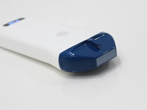
Superficial access and placement · 5P
For autologous fistulas that sit shallow with adequate calibre, mainly for pre-procedure assessment and puncture guidance, and also for CVC and dialysis catheter insertion. Representative model 5P high-frequency linear wireless probe: 10.0–14.0 MHz, four depth settings of 20 / 30 / 40 / 55 mm, in-plane and out-of-plane guidance modes.
Trade-off: this probe's puncture assistance is the simplest of the four wordings, with no automatic vessel measurement and no needle-tip enhancement. What it saves is money and weight, not precision — 10.0–14.0 MHz is sufficient at this depth band.
See this model's specifications →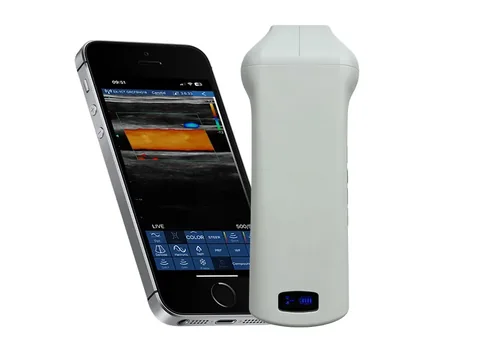
General workhorse · 6N, one probe throughout
Where pre-procedure assessment, puncture guidance and follow-up all have to be done and one probe should cover the whole pathway. Representative model 6N high-specification linear probe: 10.0–13.2 MHz, depth adjustable over 20–60 mm, 32 channels and 192 elements, all three puncture assistance items (guide line, automatic vessel measurement, needle-tip echo enhancement), 3 hours of battery life with wireless charging at any time.
Trade-off: the depth ceiling is 60 mm. Enough for autologous access; for deep follow-up on a graft or an obese patient it falls short, and you move to the 20–100 mm tier.
See this model's specifications →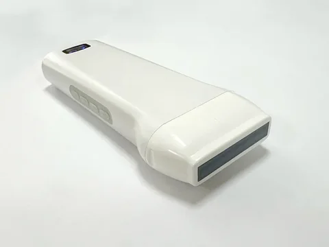
Deep access and difficult puncture · 6X
For grafts, fistulas sitting deeper, and patients with a history of difficult puncture, where you need to see beyond 60 mm and want more cues about needle position. Representative model 6X linear probe with magnetic navigation: 7.5–10.0 MHz, depth adjustable over 20–100 mm, puncture assistance covering "magnetic navigation display of needle displacement and angle, in-plane puncture guide line, out-of-plane puncture guidance and automatic vessel measurement".
Trade-off: frequency drops to 7.5–10.0 MHz in exchange for 100 mm of depth, and wall detail is not that of the 16–20 MHz tier. If a department works almost entirely on superficial autologous access, the extra depth here goes unused.
See this model's specifications →One more situation deserves separate thought: in the same dialysis centre the people doing access assessment and the people doing catheter placement are often not the same team. If the nursing group needs its own probe for PICC and dialysis catheter insertion while the medical group needs one for access follow-up, choosing two probes to their own depth requirements costs less than one "does everything" compromise — and the usual outcome of a compromise is that both ends fall short.
Five things that are easily overlooked
- "Automatic vessel measurement" is a function, not a conclusion. The parameter table reads "out-of-plane puncture guidance and automatic vessel measurement", meaning the software can measure things such as calibre. It gives a number, not a diagnosis — how much stenosis is significant and whether to intervene is a clinical judgement. The device does not carry that layer.
- The four wordings of puncture assistance differ in feel. With a guide line alone the operator has to judge on screen where the tip has reached; with needle-tip enhancement the tip stands out more in the image; with magnetic navigation the screen also gives the needle's displacement and angle. The four correspond to three different working habits — not "higher specification is better" but "who is using it and what are they used to".
- Battery figures are quoted on different bases. The 6N and 6X state "3 hours, and wireless charging at any time extends working time indefinitely"; most of the 7 series state 1.5 hours; the 9L states "2 batteries supplied, replaceable, each good for 1.5 hours". A dialysis centre runs dozens of cases a day, so the charging method has to be costed into the configuration — the hour figure alone is not enough.
- Wireless connection replaces the probe cable, not everything. The parameter tables state "wifi wireless connection", and the benefit is no cable tugging at the hand during a procedure; but each device needs a phone or tablet, so several probes in use at once means several terminals — something to think about when drawing up the rota.
- Screen position dictates puncture posture. Access puncture is done at the bedside, on a chair, with the operator's line of sight and hand position both tight. The height at which the phone is mounted and the direction it faces directly affect whether you can watch the screen and advance the needle at the same time. This cannot be found in a parameter table; it has to be tried on site.
Four lines that cannot be crossed
This section matters more than every specification above.
- Quantitative grading of stenosis is not in the device specifications. The parameter tables for this group list automatic vessel measurement, but no quantitative stenosis indices, no grading criteria and no agreement data against a reference standard. A narrowed lumen on screen is an imaging finding; whether it is a significant stenosis requires flow, pressure and clinical findings taken together.
- Access type is not distinguished in the parameter tables. Autologous arteriovenous fistula, arteriovenous graft and central venous catheter differ in depth and in what follow-up focuses on, yet our parameter tables use the single heading "vascular access" without subdividing by type. Judge depth requirements from the main access type in your own centre; do not rely on the word "applicable" alone.
- Brackets and sterile accessories need separate confirmation. Access puncture is an invasive procedure, the probe needs a sterile barrier, and some procedures also need a puncture bracket. Our parameter tables do not list bracket compatibility for each model; this page names no specific compatible model, and it has to be confirmed with the product department.
- Check the registration certificate's scope model by model. Which regions and which uses a given probe is registered for is governed by what the certificate states. Access-related procedures involve endovascular intervention and dialysis management, so obtain the original certificate before purchase and check the "intended use" column — do not substitute the "applicable departments" line from a product page.
One more point: this page discusses the selection of ultrasound imaging equipment for vascular access settings. Access function assessment also involves haemodynamics, pressure monitoring and other means, and dialysis quality management follows its own body of standards; those are outside the scope of this page and should not be mixed into the same comparison table as the ultrasound configuration.
Common questions
Q: A fistula vessel is only a few millimetres under the skin — will any linear probe do?
A: A shallow position does not mean lower requirements. Three things have to be checked: can it measure calibre (not every model has automatic vessel measurement), is the tip clear enough (needle-tip enhancement is on some models only), and is the depth setting right. The 10.0–14.0 MHz band does suit superficial work, but the assistance functions differ widely — the 5P has a guide line only, the 6N has all three.
Q: The 7H is 16–20 MHz, the highest specification — why not recommend it for everything?
A: Because its depth is only 10–40 mm. The higher the frequency, the shallower the penetration; that is a physical limit, not a configuration trade-off. The 7H shows wall layers in the finest detail within 40 mm and suits detailed observation of superficial autologous access; once the patient is obese or the access sits deeper, it simply sees nothing. Taking the highest-frequency probe to every patient is the commonest mistake in access ultrasound.
Q: How does the 6X's magnetic navigation work?
A: The wording in our parameter table is "magnetic navigation display of needle displacement and angle". It states what the function displays, not how it is implemented. That is enough for selection purposes: it gives cues about needle displacement and angle; it is not automatic puncture, and it does not replace the operator's judgement of the image.
Q: The 9L has 64 channels — is it better than the 6N's 32?
A: Channel count affects how much information the image carries; puncture assistance is a separate matter. The 9L has 64 channels, 20–100 mm of depth and two replaceable batteries, but its puncture assistance states only "in-plane puncture guide function, out-of-plane puncture guide line function", with no automatic vessel measurement and no needle-tip enhancement; the 6N has 32 channels and all three assistance items. "A clearer image" and "more cues while puncturing" are two different directions.
Q: How does this page divide work with "nursing and vascular access" and "intervention"?
A: The nursing page covers getting the tube in — finding the vessel, entering with the needle, how a nurse learns it; the intervention page covers the four technical routes to making the needle visible; this page covers how to choose the access before the procedure and how to follow it up afterwards. The probes overlap across the three pages, but the questions differ, so read them by question rather than by model.
Related reading
Nursing and vascular access: from PICC puncture to removal assessment
The practical side of access placement is on that page, including choosing in-plane or out-of-plane.
See the scenario →Urology: separating residual urine measurement from structural imaging
The other half of nephrology work sits on the urology page, where the line between dialysis and residual urine is drawn.
See the scenario →Intervention: how the needle is seen, and how to choose among four routes
The principles and limits of puncture assistance are broken down further on that page.
See the scenario →5P high-frequency linear wireless probe: 10.0–14.0 MHz
The probe quoted several times on this page; its product page gives the full four depth settings and guidance modes.
See the specifications →List your access type and usual depths and the probe answers itself
Tell us three things: which access type you mainly work on, what proportion of your patients are obese, and how the workload splits between follow-up and puncture. From those we can set the depth tier and decide whether puncture assistance is needed; you can also download the selection table and compare for yourself first.
Download the selection table See other scenarios →The specifications described here are taken from the parameter tables published on this site. Their scope is reference information based on technical specifications; the content stated in the product registration certificate prevails. For contraindications and precautions, refer to the instructions for use. The product must be operated by a licensed physician with the appropriate qualifications.

