
Obstetric and gynaecological ultrasound: why almost every service needs an endocavity probe
A gynaecology clinic mixes two quite different kinds of examination: some are about looking, others about measuring. Looking covers the uterus and adnexa, the gestational sac, the fetus and the breast; measuring covers follicle size, sac diameter, crown-rump length, biparietal diameter and femur length — figures whose value depends on being comparable. The two make opposite demands on a probe: seeing deep structures needs low frequency and penetration, measuring small structures needs mid-to-high frequency at close range. That is why a gynaecology department rarely equips itself with a single convex probe.
Four constraints to work through first
The order matters: confirm that you can see, then discuss whether you can measure accurately.
The target sits far from the skin
Uterus and adnexa lie deep in the pelvis. A transabdominal scan has to cross several layers of tissue and relies on a full bladder as an acoustic window. An endocavity probe puts the transducer closer, which is what makes the same structures readable.
What is wanted is comparable numbers
Follicle measurement, sac diameter, crown-rump length, biparietal diameter, femur length — these have to be comparable across repeat visits in the same patient. That means both measurement tools on the device and, each time, the same machine under the same conditions.
The clinic also scans breasts
Gynaecology clinics often handle breast and thyroid as well. Those are superficial structures needing a mid-to-high-frequency linear array; a convex probe cannot force itself into that and an endocavity probe is the wrong shape entirely. A device with two active faces avoids swapping back and forth.
Covers and disinfection are non-negotiable
An endocavity probe contacts mucosa, so the routine for single-use covers, gel and probe cleaning has to be fixed. This does not affect image quality, but it does affect whether the device can be used in compliance at all.
Which probe for each of the routine clinic examinations
The frequencies and depths below come from the specification tables for the wireless probes and bladder scanners on this site; each can be checked against the product page.
| Examination | Probe type | Frequency · penetration depth |
|---|---|---|
| Uterus and adnexa (transabdominal) | Convex | 3.2–5.0 MHz 90 / 160 / 220 / 305 mm |
| Uterus and adnexa, follicle tracking (endocavity) | Endocavity | 6.0–8.0 MHz 30 / 60 / 90 / 120 mm |
| Early pregnancy sac, fetal heart (close range) | Endocavity | 6.6–8.0 MHz 20–100 mm |
| Second and third trimester growth, amniotic fluid, placenta | Convex | 3.2–5.0 MHz 90 / 160 / 220 / 305 mm |
| Postpartum pelvic floor and pelvic review | Convex / endocavity | 3.2–5.0 / 6.0–8.0 MHz up to 305 mm / 30–120 mm |
| Postpartum post-void residual | Bladder scanner | 2.6 MHz or 5 MHz four-dimensional scanning measurement |
| Breast and thyroid (combined clinic items) | Linear | 7.5–10.0 MHz 20 / 30 / 40 / 55 mm |
One row deserves separate comment: the endocavity probe stops at 120 mm, while the convex reaches 305 mm. This is not because the endocavity probe is weaker, but because it does not need the depth — the transducer is already next to the target, and spare range only costs resolution. In the other direction, using it for fetal position and amniotic fluid in later pregnancy does not reach far enough. The two probes divide the work; they do not replace each other.
Another row is postpartum post-void residual. This is easily overlooked in obstetrics: postpartum voiding dysfunction needs objective residual volumes to support management decisions, and measuring with a convex probe means the operator estimating three bladder diameters and applying a formula. A bladder scanner takes a different route — four-dimensional scanning measures the volume directly, and the probe is a single handheld unit with no mainframe required.
Three common configurations, chosen by the clinic's actual caseload
The three tiers correspond to three workloads; they are not a cheaper-to-better ladder.
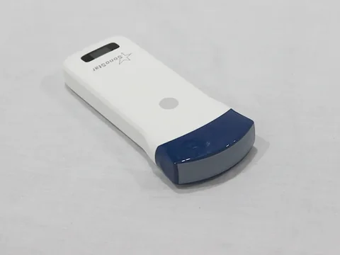
Transabdominal only · Starting with one convex probe
For clinics whose work is mainly health checks, second and third trimester scanning and routine gynaecological screening. Representative model: the 5C convex wireless ultrasound probe — 3.2–5.0 MHz, penetration depth 90 / 160 / 220 / 305 mm, with measurement tools for crown-rump length, biparietal diameter, sac diameter, femur length, head circumference, abdominal circumference and estimated fetal weight.
The trade-off is clear: the resolution of the endocavity tier and the close-range advantage in early pregnancy are given up, in exchange for one probe covering abdominal, obstetric and urological work. Transabdominal examination requires a full bladder, and that belongs in the appointment reminder.
See the specifications →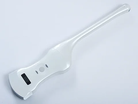
Transabdominal plus endocavity · One device covers both
For institutions doing both gynaecology and obstetrics at moderate volume, this is the least troublesome dividing line. Representative model: the 5CT convex plus endocavity wireless ultrasound probe — convex 3.2–5.0 MHz, depth 90 / 160 / 220 / 305 mm; endocavity 6.0–8.0 MHz, depth 30 / 60 / 90 / 120 mm.
One probe, two active faces, switched with a button. Early pregnancy and follicle tracking on the endocavity face, later pregnancy and abdomen on the convex, with no changing of parts or machines in between. Most obstetrics and gynaecology departments in secondary hospitals settle at this tier.
See the specifications →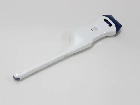
Endocavity-led · Adding the superficial half
For fertility centres, infertility clinics and services that also scan the breast. Representative model: the 5LT linear plus endocavity dual-head probe — linear 7.5–10.0 MHz, depth 20 / 30 / 40 / 55 mm; endocavity 6.0–8.0 MHz, depth 30 / 60 / 90 / 120 mm, 128 elements and 32 channels.
The endocavity face handles follicles, uterus and adnexa; the linear face handles breast, thyroid and superficial nodes. The cost is no low-frequency convex, so deep structures in later pregnancy are out of range and need separate provision.
See the specifications →A lighter option still: a single endocavity-only probe, the 5T (6.6–8.0 MHz, penetration depth 20–100 mm, 128 elements, 32 channels, R13), for a dedicated follicle-tracking station in a fertility centre. Its range is short but mid-frequency, so follicle borders close in come out clearer than with a convex probe. The drawback is that beyond 100 mm it sees nothing, and abdominal and obstetric work is beyond it.
Where volume is high and deeper abdominal scanning is needed, move up within the convex tier: the 7C (3.2–5.0 MHz, depth 90 / 160 / 220 / 305 mm, 192 elements, R60) or the 8C (3.2–5.0 MHz, depth 90 / 160 / 240 / 300 mm). The depth settings differ; choose against the depths you actually work at rather than guessing from the model number.
Four things that often get missed
- Covers are not optional. The endocavity probe contacts mucosa, so single-use covers, sterile gel and post-examination cleaning have to form a fixed routine. Draw up the consumables list at the purchasing stage; it is far less trouble than filling the gap after installation.
- Preparation decides the image. Transabdominal scanning needs a full bladder as an acoustic window; endocavity scanning needs an empty one. On the same device, getting preparation right or wrong can make more difference than changing machines. That sentence belongs in the appointment message, not only on the department wall.
- Comparability in follow-up comes from identical conditions. For follicle tracking and sac follow-up, repeat examinations in the same patient have to be comparable. The method is plain: use the same device, the same measurement plane and the same measuring habits where possible, and note them in the report. The device supplies the measurement tools; comparability comes from the protocol.
- Do not expect one convex probe to cover the postpartum pelvic floor. A convex probe can show the pelvis and residual urine, but systematic assessment of the pelvic floor musculature and supporting structures is a specialty of its own, needing its own examination protocol and interpretive experience. Being clear before purchase about how far you can go is better than finding the gap afterwards.
Three lines not to cross
This section matters more than every specification above it.
- One scan does not close a diagnosis. Confirming an early pregnancy, judging whether an adnexal mass is benign or malignant, excluding an ectopic pregnancy — none of these can be concluded from a single image. They require history, laboratory tests and follow-up. The device supplies images and measurements; the judgement belongs to the physician.
- Fetal structural screening is not work for a portable device. Systematic fetal anomaly screening has defined gestational windows, view protocols and operator qualification requirements. A portable device can follow fetal heart, amniotic fluid, presentation and growth measurements, but it does not replace screening carried out by a qualified institution. In primary care this especially belongs in the referral protocol.
- Measurements depend on correct planes, not on how automatic the device is. Whether head circumference, abdominal circumference and femur length are comparable depends on being measured in the standard plane. The device provides the tools and the arithmetic; whether the plane was correct remains the operator's responsibility.
One further point: alongside ultrasound, an obstetrics department also uses equipment of an entirely different kind, such as fetal monitoring. This page deals with ultrasound imaging equipment only. The selection and interpretation standards for fetal monitoring are outside its scope, and the two should not be compared in the same configuration table.
Common questions
Q: What exactly is the difference between transabdominal and endocavity?
A: It comes down to the distance from transducer to target. Transabdominal scanning crosses several layers of tissue and relies on a full bladder as an acoustic window, hence low-frequency convex at 3.2–5.0 MHz with depth to 305 mm. Endocavity scanning places the transducer closer, so mid-frequency 6.0–8.0 MHz at 30–120 mm is enough, with better close-range resolution. A device carrying both faces can switch by examination type without a second machine.
Q: If we only do health checks and pregnancy or IUD checks, do we need an endocavity probe?
A: Look at the actual caseload first. If it is transabdominal routine screening and later-pregnancy follow-up only, one convex probe is enough, and the budget is better spent on depth and measurement tools. If early pregnancy, follicle tracking and endometrial assessment make up a fair share, the endocavity tier shows its advantage at once — done transabdominally, those examinations require the patient to hold a full bladder and the image is more affected by body habitus.
Q: Which probe in early pregnancy?
A: In early pregnancy the structures are small and sit deep, so the close-range advantage of an endocavity probe is at its clearest. Transabdominal scanning can do it too, but requires a well-filled bladder and is more affected by habitus and bowel gas. In practice many services work endocavity first, with transabdominal as the fallback.
Q: Can one ultrasound device cover both the postpartum pelvic floor and residual urine?
A: Residual urine, yes; the pelvic floor needs separate treatment. Residual volume can be estimated with a convex probe, or measured directly with a dedicated bladder scanner (the BProbe-2 at 2.6 MHz and the BProbe-5 at 5 MHz, both using four-dimensional scanning). Systematic assessment of pelvic floor musculature and supporting structures is another field, requiring its own protocol.
Q: Can the breast be scanned on the same device?
A: Yes, but it needs a linear face. Breast and thyroid are superficial structures requiring the 7.5–10.0 MHz linear range at 20–55 mm depth; neither convex nor endocavity suits them. A model with a linear face (the 5LT, for example) covers both, while convex-only or endocavity-only models cannot.
Related reading
Using portable ultrasound well without a dedicated sonographer
Who operates, how they are trained, when referral is mandatory — gynaecological screening runs into the same three questions.
Read →Handheld, portable or trolley ultrasound: start with where it will sit
Form factor before specifications — where clinic and ward divide is set out here.
Read →Primary care screening: working back from a day's caseload
If gynaecology is only one item in the clinic, start with the approach on this page.
See the scenario →Bladder scanners: two technical routes to postpartum residual urine
The difference between convex estimation and four-dimensional measurement can be compared model by model in the product section.
See the product section →List the examinations and the configuration follows
A maternal and child health hospital, the obstetrics department of a general hospital, a fertility centre and a community health centre have very different caseload structures. The Selection Guide section of this site carries a comparison table arranged by examination site and penetration depth, for discussion within the department.
Download the selection guide See other scenarios →The specifications in this article are taken from the product specification tables on this site. This document is a reference explanation based on technical parameters; where it differs, the content stated on the product registration certificate takes precedence. For contraindications and precautions, see the product instructions for use. The product must be operated by a licensed physician with the appropriate qualifications.

