
Ultrasound for primary care clinics: work backwards from a day's caseload
Township health centres, community health service centres and village clinics buying ultrasound tend to go wrong in one of two ways. One is copying the equipment list of the hospital above them and ordering a trolley system, only to find there is no dedicated sonographer, reports do not get written, and the machine ends up in a cupboard. The other is picking a cheap single probe that cannot reach deep enough for the abdomen and cannot resolve anything superficial either, so it is set aside after two uses. This page is not about brands. It is about one thing: work out three numbers first, then decide how many units, in what form factor, and whether endocavity and cardiac modes are needed.
Three hard constraints that come with this setting
The same device that feels natural in an ultrasound department may not feel natural in a primary care clinic. These three points are why.
No dedicated sonographer
In most clinics a clinician scans alongside their own caseload — a dozen or so patients a day, with image-optimisation skills still being built up. The device needs to work the moment it is switched on. Preset examination types and automatic measurement are worth more here than an extra row of knobs.
The room and the power supply keep changing
Village screening days, home visits, public-square clinics: a socket may not be there, and a network even less so. Whether the device can work away from its usual environment decides whether it is equipment you can take out, or equipment that only works in one room.
A mixed caseload, and one probe is not enough
Liver, gallbladder, pancreas, spleen and kidneys in the morning; thyroid and carotid in the afternoon; two post-void residual checks in between. That spread is too wide for a single-frequency probe — one end of it always comes out wrong.
What you scan, which probe, and how deep
The table below lines up examination site with probe type, frequency and penetration depth. The figures are taken from the product specification tables on this site, so they can be checked item by item.
| Examination | Probe type | Frequency | Penetration depth |
|---|---|---|---|
| Liver, gallbladder, pancreas, spleen, kidneys; ascites | Convex | 3.2–5.0 MHz | 90 / 160 / 220 / 305 mm |
| Thyroid, breast, superficial masses | Linear | 7.5–10.0 MHz | 20 / 40 / 60 / 80 mm |
| Carotid, lower-limb vessels | Linear | 7.5–10.0 MHz | 20 / 40 / 60 / 80 mm |
| Uterus and adnexa (transabdominal), post-void residual, prostate | Convex | 3.2–5.0 MHz | 90 / 160 / 220 / 305 mm |
| Basic cardiac screening (cardiac mode) | Cardiac mode | 3.2–5.0 MHz | 90 / 120 / 140 / 160 mm |
| Basic cardiac screening (phased array) | Phased array | 2.2–3.6 MHz | 90 / 160 / 200 / 240 mm |
| Gynaecological endocavity examination | Endocavity | 6.0–8.0 MHz | 30 / 60 / 90 / 120 mm |
The line worth remembering in this table is the boundary between abdominal and superficial work: a convex array has to see more than ten centimetres down, a linear array only two or three. Covering both ends with one device in a primary care setting means choosing a dual-head or triple-in-one probe that can switch modes, not stretching a single probe across every examination.
Three tiers by daily caseload
These three tiers are not a cheaper-to-better ladder; they correspond to three different workloads. Work backwards from an actual day, not from a budget.
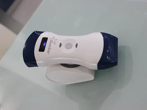
Entry tier · One dual-head probe for abdomen and superficial work
For village clinics and community stations scanning fewer than 10 patients a day, with only one or two clinicians able to operate. Representative model: the 5CL dual-head wireless ultrasound probe — convex 3.2–5.0 MHz, penetration depth 90 / 160 / 220 / 305 mm; linear 7.5–10.0 MHz, penetration depth 20 / 40 / 60 / 80 mm. One probe with two active faces covers abdomen, thyroid and carotid without changing anything mid-session.
The connection is wireless: the probe emits its own WiFi signal and a phone or tablet receives the image directly, so whether the room has a network makes no difference to use.
See the specifications →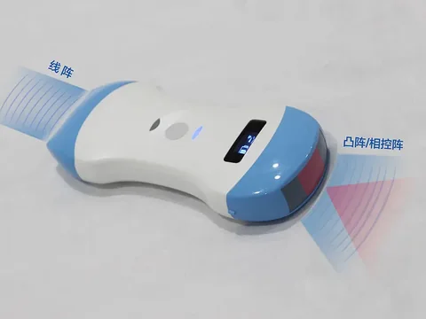
Main tier · Adding a cardiac mode
For township health centres scanning 10–30 patients a day that also need basic cardiac and vascular screening. Representative model: the 5CPL triple-in-one wireless ultrasound probe — convex and cardiac mode 3.2–5.0 MHz, linear 7.5–10.0 MHz; the three penetration depth sets are convex 90 / 160 / 220 / 305 mm, cardiac mode 90 / 120 / 140 / 160 mm, linear 20 / 40 / 60 / 80 mm. The 6CPL shares the same structure; settle on it against the actual quotation and accessories.
The extra cardiac mode is there to show whether there is pericardial fluid and roughly what the chamber proportions look like. It is not a substitute for a dedicated cardiac study in an ultrasound department.
See the specifications →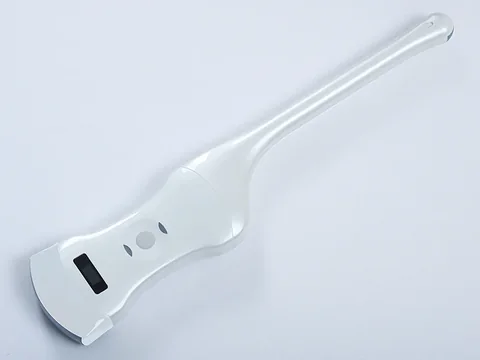
Add-on tier · When gynaecology passes about a third of the caseload
Transabdominal views of the uterus and adnexa often come out poorly in patients with bowel gas or a larger body habitus, and this is where an endocavity probe fills the gap. Representative model: the 5CT convex plus endocavity wireless ultrasound probe — convex 3.2–5.0 MHz, depth 90 / 160 / 220 / 305 mm; endocavity 6.0–8.0 MHz, depth 30 / 60 / 90 / 120 mm.
Endocavity examination is an invasive procedure. It should be carried out by personnel with the appropriate qualifications, following the protocol of the institution concerned, with proper probe covering and disinfection.
See the specifications →Three things that get overlooked and cannot be recovered later
- Images that can be stored and sent on. The real value in primary care is follow-up: measuring the same nodule again three months later only means something if last time's record is there to compare against. Before buying, confirm the images can be kept on a phone, exported as files, and archived to a computer or system in line with institutional rules.
- Cleaning, disinfection and gel between patients. Probe covers, disinfectant wipes and coupling gel are consumables, not accessories, and belong in the running-cost calculation at the purchasing stage. Endocavity probes additionally need isolation covers, in line with the infection-control requirements of the institution.
- Who reads the images. Buying the device is only the first step; after that someone has to mentor and someone has to review. Write down the situations that must be referred as a written policy for the institution and put it on the wall of the examination room — it holds up better than word of mouth.
The first and third of these never appear on a quotation, yet they decide whether the equipment sits idle. Before ordering, ask one question: in six months' time, whose hands will this device be in, and what will it be used for? If the answer is clear, place the order.
Common questions
Q: If I only scan a dozen patients a day, am I buying something too big?
A: The criterion is not the patient count but whether anyone operates it on a fixed basis. Low volume with a designated operator: one dual-head portable device is enough. Low volume with no designated operator: a trolley system is more likely to sit idle, because moving and setting it up costs more effort, and a busy clinician will not bother.
Q: With no dedicated sonographer, how should equipment and training be matched?
A: Start with equipment that has a short operating path — preset examination types, automatic measurement and guide lines cut down the time spent adjusting the image. Then schedule training before installation: learn the two high-frequency examinations, abdomen and thyroid, first, and pick up the rest as you go. However well designed the device is, nobody becomes competent without someone showing them.
Q: Is one dual-head probe enough, or do I need several?
A: Two numbers decide it: what share is abdominal versus superficial, and whether basic cardiac screening is needed. If abdomen plus superficial work makes up nine tenths and there is no cardiac work, a dual-head is enough. Only when basic cardiac screening is a routine part of the caseload does a cardiac-mode or phased-array model come into consideration.
Q: The village clinic has no network. Can the device still be used?
A: Yes. On the wireless models the probe emits its own WiFi signal and the phone or tablet connects to the probe directly to take the image; it does not depend on the setting having a network. What does need a connection is sending the records on afterwards, and that step can wait until you are somewhere with one.
Q: Do I need colour Doppler?
A: It depends on the examinations. For thyroid, breast and carotid — superficial and vascular work — colour flow helps the assessment. For routine abdominal work and post-void residual only, B-mode plus measurement is sufficient. The wireless probe series on this site offers B, B/M, Color, PDI and PW imaging modes; check the specification table for the exact configuration of a given model.
Related reading
Equipping a primary care ultrasound service: working back from a day's clinic volume
The full worked version of the same approach, with caseload, examination mix and staffing laid out as three variables.
Read →Using portable ultrasound well without a dedicated sonographer
What to do when the device arrives and nobody knows how to use it: positioning, what can be scanned, and when referral is mandatory.
Read →What to take on a rural outreach clinic: a trade-off list for portable ultrasound
Power, network and handling — the three things that decide whether the day gets off the ground.
Read →Handheld, portable or trolley ultrasound: start with where it will sit
Settle the location first, then discuss specifications. Get the form factor wrong and everything after it is skewed.
Read →Read the table first, then decide which unit
The Selection Guide section of this site carries a wireless probe comparison table, listing models by examination site and penetration depth. It can be downloaded and printed for departmental discussion and pre-purchase configuration checks.
Download the selection guideThe specifications in this article are taken from the product specification tables on this site. This document is a reference explanation based on technical parameters; where it differs, the content stated on the product registration certificate takes precedence. For contraindications and precautions, see the product instructions for use. The product must be operated by a licensed physician with the appropriate qualifications.

