
Cardiovascular ultrasound: a probe can answer whether, only a console can answer how much
In cardiac work the commonest selection error is to run two things together. One is whether — is there effusion, has a chamber enlarged, is a valve moving properly. The other is how much — what is the ejection fraction, what is the cardiac output, what is the transvalvular gradient. The first a probe can answer on its own; the second needs measurement and analysis software on a mainframe. The range on this site draws that line clearly: the wireless phased arrays stop at continuous-wave Doppler, while ejection fraction, left ventricular volume and cardiac output appear only in the cardiac function and vascular analysis of a console. This page follows that line.
Four constraints to work through first
The order matters: decide first whether the question is whether or how much.
The heart sits behind the ribs
The heart is enclosed by sternum and ribs, and the acoustic window is a few intercostal spaces. Getting through takes a low-frequency phased array: the 5PA and 6PA here are both 2.2–3.6 MHz with an 80-degree sector and depth settings running from 90 mm to 240 mm. Physics decides this, not configuration preference.
Low and high velocity are two different techniques
Structure is shown by two-dimensional imaging and colour; blood velocity needs Doppler. Pulsed wave (PW) gives depth selection but cannot measure high velocities; continuous wave (CW) measures high velocities but has no depth resolution. The modes listed for the 5PA stop at PW, while the 6PA's explicitly include CW — and that is the most substantive difference between the two probes.
Functional quantification lives on the mainframe
Ejection fraction, left ventricular volume, cardiac output, pressure half-time, transvalvular gradient and Qp/Qs appear on this site only in the cardiac function and vascular analysis of the C200. What a probe provides is images and basic measurements — distance, area, circumference, heart rate — and the two are not the same level.
A conclusion is needed at the bedside
On a cardiology ward round, in the emergency department, in the ICU, during anaesthetic induction, the device has to reach the bedside and produce an image from switch-on. That puts the question of form factor first: for whether at the bedside, a wireless probe is the whole device and connects to a tablet; reporting and quantification are what bring you back to a console.
Ask what has to be answered, then match the configuration
The figures below come from the specification tables for the 5PA, 6PA, 5LPA, C200 and C600 on this site.
| Question to be answered | Configuration level | Key figures |
|---|---|---|
| Is there effusion; chamber size; volume status | 5PA phased-array probe | 2.2–3.6 MHz · up to 240 mm B, B/M, Color, PDI, PW |
| High-velocity flow (continuous wave) | 6PA phased-array probe | 2.2–3.6 MHz · includes CW 192 elements · up to 200 mm |
| Cardiac and vascular access together | 5LPA dual-head probe | phased array 3.3–3.6 MHz / linear 7.5–10.0 MHz R80 / L25 |
| Ejection fraction, cardiac output, transvalvular gradient | C200 console | PWD standard, CWD optional PWD ≥6 m/s · CWD ≥15 m/s |
| Tissue Doppler, anatomical M-mode | C600 console | B, C, PW, CW, BCD triplex, TDI real-time wide view, anatomical M-mode |
The dividing line in this table falls at the fourth row. The first three rows depend on a probe; the last two on a mainframe. This is not marketing language but the boundary of the specification tables themselves: the imaging modes of the 5PA and 6PA go no further than CW, and their measurement functions list distance, area, circumference and heart rate; whereas under cardiac function and vascular analysis on the C200 there are ten items side by side — ejection fraction, left ventricular volume, cardiac output, pressure half-time, transvalvular gradient, Qp/Qs, velocity time integral, body surface area, ICA/CCA diameter ratio and stenosis percentage.
So the answer to a quick look at the bedside and the answer to producing a report should not be the same device. The first wants a fast start and an adequate image; the second wants a full measurement set and a printed report with images. Force the two onto one machine and it is either needlessly expensive or found wanting when the report is written.
One more row deserves separate comment: the 5LPA is a phased-array plus linear dual head. The phased array at 3.3–3.6 MHz with 90–240 mm depth covers the heart; the linear at 7.5–10.0 MHz with 20–100 mm covers vascular access. It is the one among these probes whose specification table explicitly lists cardiac PW-mode VTI measurement and BM-mode LVID measurement, and it adds needle-tip enhancement. To do a heart study and then look at vascular access on the same device, this is the convenient choice.
Three common configurations, chosen by how far the work goes
The three tiers are not about price but about whether the work stops at the image or goes on to quantification.
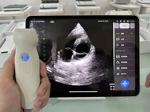
Tier one · 5PA: structure and volume status
Mainly for rapid bedside assessment: is there pericardial fluid, are the chambers enlarged, is the patient volume-replete. Representative model: the 5PA phased-array wireless ultrasound probe — 2.2–3.6 MHz, 128 elements, 32 channels, 80-degree sector, depths of 90 / 160 / 200 / 240 mm, modes B, B/M, Color, PDI and PW.
The trade-off: the modes stop at PW, with no CW, so high-velocity flow cannot be measured; battery life is 1 hour with wired charging, so consecutive cases need charging provision. What it buys is 120 g held in one hand, and an image within a minute at the bedside.
See the specifications →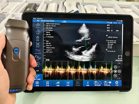
Tier two · 6PA: adding CW
Where high-velocity flow and continuous-wave Doppler are needed. Representative model: the 6PA phased-array wireless ultrasound probe — 2.2–3.6 MHz, 192 elements, 32 channels, 80-degree sector, depths of 90 / 130 / 160 / 200 mm, modes including CW, gain 30–105 dB, 2600 mAh battery and 2 hours of use.
The trade-off: elements rise from 128 to 192, maximum depth comes in from 240 mm to 200 mm, and battery life doubles. For high-velocity flow at the bedside this tier is necessary; for structure alone, the extra is not used.
See the specifications →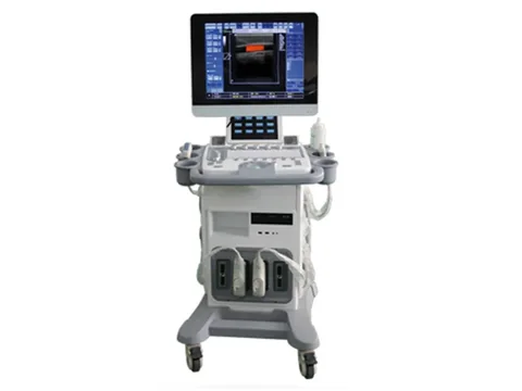
Tier three · C200: going on to quantification
Where ejection fraction is to be produced, cardiac output calculated and an illustrated report written. Representative model: the C200 trolley system — 15-inch touchscreen, image depth 2–30 cm, frequency 2–10 MHz, PWD standard and CWD optional, maximum measurable velocity ≥6 m/s unidirectional on PWD and ≥15 m/s unidirectional on CWD, with the phased-array probe a separate option.
The trade-off: a console takes floor space, needs power, and needs someone who knows its measurement menus. What it buys is the whole cardiac function and vascular analysis group — ejection fraction, left ventricular volume, cardiac output, pressure half-time, transvalvular gradient and Qp/Qs. A probe cannot provide that.
See the specifications →One more combination is worth knowing: the 5LPA (phased array 3.3–3.6 MHz plus linear 7.5–10.0 MHz, 128 elements, R80 / L25, 230 g). Its phased-array section carries cardiac PW-mode VTI measurement and BM-mode LVID measurement, the linear section covers vascular access and puncture guidance, and the specification table lists needle-tip enhancement. One device doing heart and access is less trouble than carrying two probes.
Tissue Doppler (TDI), anatomical M-mode and real-time wide-view imaging mean going to the C600: its imaging modes are listed as B, C, PW, CW, BCD triplex, TDI, real-time wide-view imaging, trapezoidal imaging (linear probes only) and anatomical M-mode. Nothing in this class exists on a probe.
Four things that often get missed
- The 5PA runs for only 1 hour. The specification says 1 hour, charged by cable. Several patients in a row at the bedside need a charging window in between, and this is easy to trip over in the emergency department and the ICU.
- The supported operating systems differ. In the 5PA and 6PA specification tables the software platform is listed as Apple iOS only; the 5LPA lists Apple iOS, Android and Windows. A department using Android tablets should confirm this first, or the software will not install on arrival.
- Maximum phased-array depth differs by model. The 5PA reaches 240 mm, the 6PA 200 mm, the 5LPA 240 mm, and the C200 console 2–30 cm. A larger number is not automatically better, but a structure beyond the maximum simply will not be seen.
- Probe angle affects the image more than probe specifications do. The cardiac windows are narrow, and where the intercostal space lies decides which views can be obtained. On the same probe, ten degrees of angulation can make more difference than changing probe. This is a matter of technique and practice.
Four lines not to cross
This section matters more than every specification above it.
- Do not ask a probe for an ejection fraction. In the specification tables on this site, ejection fraction, left ventricular volume and cardiac output appear only under cardiac function and vascular analysis on the C200. The measurement functions of the wireless probes list distance, area, circumference and heart rate. Attributing quantitative indices to a probe is the claim most likely to be challenged on the spot by an experienced user.
- Whether and how much are two clinical questions. Seeing pericardial fluid at the bedside answers whether; how much fluid, and whether it is causing tamponade, are judged alongside other indices. The device provides images; the judgement belongs to the physician.
- A single bedside study does not replace a full cardiac examination. Bedside assessment has limited views and limited time and serves rapid screening and serial observation; systematic assessment of cardiac structure and function needs a complete protocol, standard views and qualified personnel.
- This page covers ultrasound only. Cardiovascular practice also uses equipment of other kinds, such as electrocardiography, which has its own product section on this site. The selection logic differs, and the two should not be compared in the same configuration table.
One further point: view standards, measurement conventions and report formats in echocardiography are set by professional bodies and by the institution concerned. What this page gives is a device-side mapping of specifications to tiers, and it does not replace any protocol.
Common questions
Q: Can a wireless phased array measure ejection fraction?
A: In the specification tables on this site, ejection fraction, left ventricular volume, cardiac output, pressure half-time, transvalvular gradient and Qp/Qs appear only in the cardiac function and vascular analysis of a console (the C200, for example). Probe measurement functions list distance, area, circumference and heart rate, plus cardiac PW-mode VTI and BM-mode LVID measurement on the 5LPA. Producing an ejection fraction means going to that console tier.
Q: What is the difference between the 5PA and the 6PA?
A: Four things. Elements: 128 against 192. Modes: the 5PA stops at PW, the 6PA includes CW. Depth settings: 90 / 160 / 200 / 240 mm on the 5PA against 90 / 130 / 160 / 200 mm on the 6PA. Battery: 1 hour against 2 hours (2600 mAh). Both are 2.2–3.6 MHz at 80 degrees. For high-velocity flow there is only the 6PA.
Q: For pericardial fluid in the emergency department, is a console needed?
A: In most cases no. The 5PA at 2.2–3.6 MHz with depth to 240 mm, in B mode and colour, is enough to answer whether there is fluid. The value of a console lies in quantification and reporting, not in whether something can be seen. The division of work at the emergency bedside is set out in more detail on the emergency and point-of-care page.
Q: The 5LPA does the work of two probes — how is it used?
A: The phased-array face (3.3–3.6 MHz, 90–240 mm) covers the heart, the linear face (7.5–10.0 MHz, 20–100 mm) covers vascular access and puncture guidance, switched by button. It is the one among these probes whose table explicitly lists cardiac PW-mode VTI and BM-mode LVID measurement, and it adds needle-tip enhancement. The cost is 230 g, nearly twice the weight of the 5PA or 6PA.
Q: When is CW essential?
A: When high-velocity flow has to be measured. Pulsed wave has depth resolution but aliases at high velocities; continuous wave has no depth resolution but reaches higher velocities. The C200 specification states it clearly: ≥6 m/s unidirectional on PWD, ≥15 m/s unidirectional on CWD. Structure does not need CW; high-velocity flow does.
Related reading
Emergency bedside: one question at a time
Pericardial fluid, free gas, whether a vessel is open — the whether side is covered here.
See the scenario →Anaesthesia and pain: configuring perioperative cardiac monitoring
The same phased array behaves differently in theatre.
See the scenario →Handheld, portable or trolley ultrasound: start with where it will sit
How bedside and examining room divide decides both form factor and budget.
Read →Selection guide: a comparison table by site and depth
Take the tier table from this page with you for departmental discussion.
See the selection guide →Decide first whether a report is needed and half the configuration is settled
Bedside assessment and a full cardiac examination are two different configurations. Tell us where it will be used, how many patients a day and who will operate it, and we can advise on those three points; or download the comparison table and work through it yourself first.
Download the selection guide See other scenarios →The specifications in this article are taken from the product specification tables on this site. This document is a reference explanation based on technical parameters; where it differs, the content stated on the product registration certificate takes precedence. For contraindications and precautions, see the product instructions for use. The product must be operated by a licensed physician with the appropriate qualifications.

