
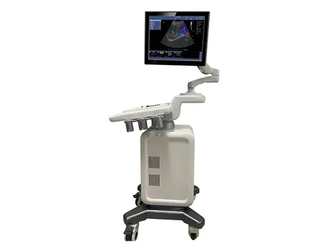
C300 Colour Doppler Ultrasound System
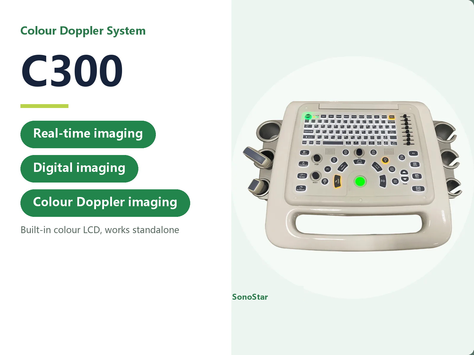
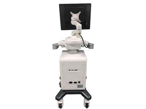
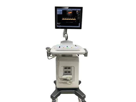
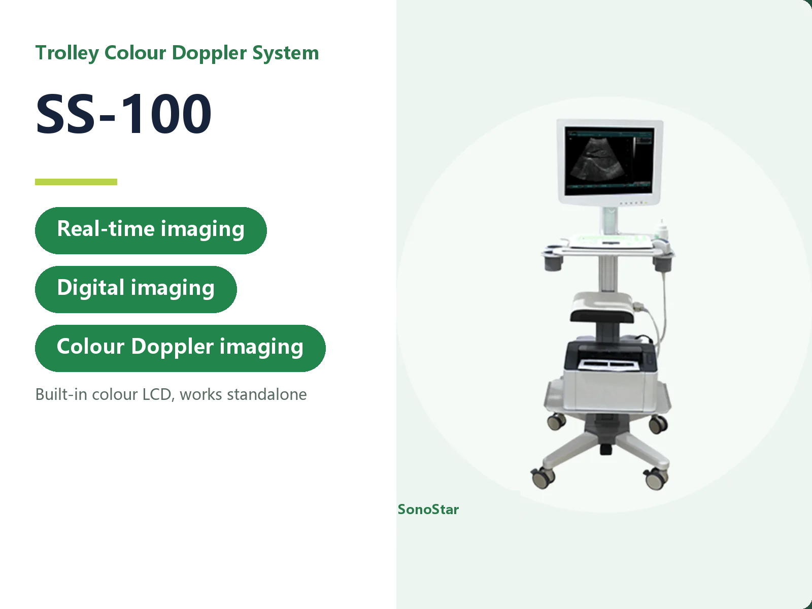
C300 trolley colour Doppler with an abdominal convex probe and built-in battery. Colour Doppler, tissue harmonic imaging and fetal growth curves.
- Brand: SonoStar
- Price: On request
- Probe frequency: Complete system with several probe connectors; a range of probes is available (see specifications)
- Connection: Self-contained colour LCD; works standalone, no external display needed
- Charging: Built-in battery supplied as standard (see specifications)
- Category: Trolley Ultrasound
Product overview
The C300 is a trolley-mounted colour Doppler ultrasound system supplied with an abdominal convex probe and a built-in battery. Linear, endocavity, microconvex and phased array probes and a needle guide are available as options, so the same trolley can cover abdominal, obstetric, vascular and superficial work.
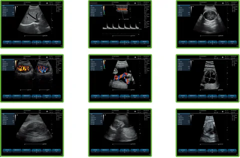
Imaging. Colour Doppler flow imaging, 2D grey-scale imaging and power Doppler imaging form the base. Tissue harmonic imaging, spatial compounding, independent deflection on the linear probe and speckle noise reduction improve contrast and reduce artifact.
Measurement. General measurements cover distance, area, perimeter, volume, area ratio, distance ratio, angle, S/D velocity, time, heart rate and acceleration. The obstetric and fetal package includes a fetal weight algorithm, growth curve display and fetal echocardiography measurements, including left ventricular function and left ventricular mass. The sample volume has at least 8 steps, and annotations can be inserted, edited and saved.
System. A 15-inch high-resolution colour LCD, zero-insertion-force metal probe connectors with two active universal ports, a 320 GB hard disk, and DICOM 3.0 output.
Technical specifications
Imaging functions
| Item | Data |
|---|---|
| Base imaging | Colour Doppler flow imaging, 2D grey-scale imaging, power Doppler imaging |
| Advanced imaging | Tissue harmonic imaging, spatial compounding, independent deflection on the linear probe, speckle noise reduction |
Measurement and analysis
| Item | Data |
|---|---|
| General measurements | Distance, area, perimeter, volume, area ratio, distance ratio, angle, S/D velocity, time, heart rate, acceleration |
| Obstetric and fetal | Fetal weight algorithm, growth curve display, fetal echocardiography measurements (including left ventricular function and left ventricular mass) |
| Vascular flow | Sample volume adjustable in at least 8 steps |
| Custom annotation | Insert, edit, save |
System
| Item | Data |
|---|---|
| Colour monitor | 15-inch high-resolution colour LCD |
| Probe connectors | Zero-insertion-force metal connectors, 2 active universal ports |
| Input | Digital signal interface |
| Output | Composite video, RGB colour video, S-video, USB |
| DICOM | DICOM 3.0 interface |
| Hard disk | 320 GB; patient still and moving images stored, managed and replayed on the unit |
Probes
| Item | Data |
|---|---|
| Multi-frequency | Variable frequency, 2.5-10.0 MHz |
| Probe types | Convex, linear, endocavity |
| Frequency steps | 5 steps per probe; fundamental and harmonic frequencies selectable |
| Abdominal probe | 2.5-6.0 MHz, maximum display depth 240 mm |
| Superficial probe | 6.5-10.0 MHz, maximum display depth 150 mm |
| B/D combined | Linear B/PWD, convex B/PWD, sector B/PWD |
| Needle guidance | Optional probe needle guide with adjustable needle line angle |
2D grey-scale
| Item | Data |
|---|---|
| Frame rate | Convex probe, full field, 19 cm depth: 55 frames per second |
| Maximum zoom | 5x |
| Transmit focus | 4 focal zones |
| Reception | Parallel multi-beam processing |
| 2D gain | Continuously adjustable |
| Maximum scanning depth | 24 cm |
| Cine memory | 256 seconds |
| Beamformer | Digital beamformer, digital full-depth dynamic focus, digital dynamic aperture and apodisation, focal position adjustable over the whole imaging field |
| Presets | Optimised per organ to reduce adjustment during scanning |
| TGC | 8 segments |
| Pseudo-colour | 6 maps |
| Sector angle | 4 adjustable steps |
| Acoustic power | Visible and adjustable |
Spectral Doppler
| Item | Data |
|---|---|
| Mode | Pulsed wave Doppler (PW) |
| Doppler frequency | PW frequency adjustable |
| Maximum velocity | 2 m/s forward or reverse flow |
| Auto envelope | Automatic Doppler envelope measurement and calculation |
| Sample width and position | Width 1-10 mm |
| Display control | Invert display (left-right, up-down) |
| Auto measurement | Real-time PW auto measurement |
| Cine playback | Automatic cine playback |
Colour Doppler
| Item | Data |
|---|---|
| Doppler gain | Continuously adjustable |
| Colour enhancement | Supported |
| Dual display | B + COLOR side by side on one screen |
| Baseline | Adjustable +/-15 steps in colour mode |
Configuration
| Item | Data |
|---|---|
| Standard | Main unit x1, abdominal convex probe x1, built-in battery x1 |
| Optional | Linear, endocavity, microconvex and phased array probes; needle guide |
Specifications updated 2026-11-01
Typical applications
The application scenarios on this page describe the device based on its technical data and configuration. The actual scope of application is the one stated in the product registration certificate. Follow your institution's procedures and the instructions for use.
| Setting / department | Typical use | Configuration note |
|---|---|---|
| Emergency / ICU | Bedside scanning of the abdomen, the heart and pleural or peritoneal fluid | Self-contained unit with a colour LCD |
| Obstetrics / gynaecology / urology | Gynaecological, obstetric and urinary tract examinations | Convex and endocavity probes available |
| Cardiology / small parts | Cardiac, peripheral vascular, superficial tissue and small organ examinations | Phased array and high-frequency linear probes available |
| Primary care / health screening | General whole-body ultrasound | Several probe connectors, configured to need |
Usage and selection notes
The C300 is a complete-system product and belongs to a different product line from the wireless probes.
A number of probe types can be fitted, so the configuration supplied is the one on the order sheet. Please confirm the selection with our product department.
For contraindications and precautions, refer to the instructions for use. The device must be operated by a licensed physician with the relevant qualifications.
The technical data on this page is taken from the product technical documentation; where it has been updated, the latest version applies. The listed departments and application scenarios describe the device based on its technical data, and the actual scope of application is the one stated in the product registration certificate. This page makes no claim about clinical outcome and does not constitute clinical guidance.
Related products
Need a quote or a sample for evaluation?
Tell us how you plan to use the system and our sales team will reply within one business day.
Get a quote
