
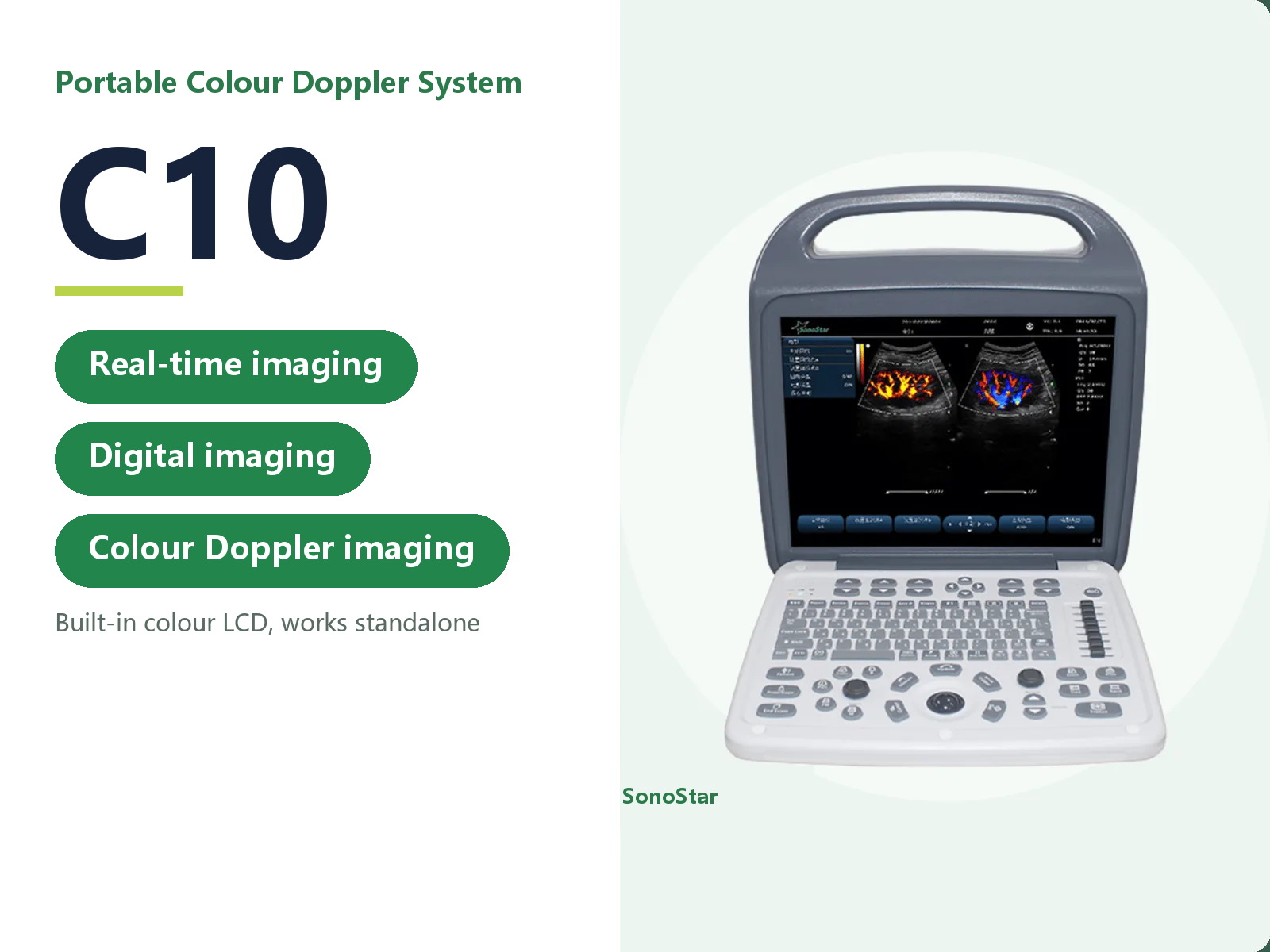
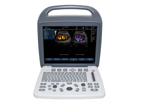
C10 Portable Colour Doppler Ultrasound System
Portable colour Doppler with a 15-inch medical LCD and two probe sockets. Covers abdomen, gynaecology, obstetrics, cardiac and paediatric work. 7.5 kg.
- Brand: SonoStar
- Price: On request
- Probe frequency: Not applicable to the main unit - this is a console with several probe sockets; see the technical data for the optional probes
- Connection: Not applicable - the console has its own colour LCD and works independently, with no external display needed
- Charging: Built-in battery, 7000 mAh or more (see technical data)
- Category: Portable Ultrasound
Product overview
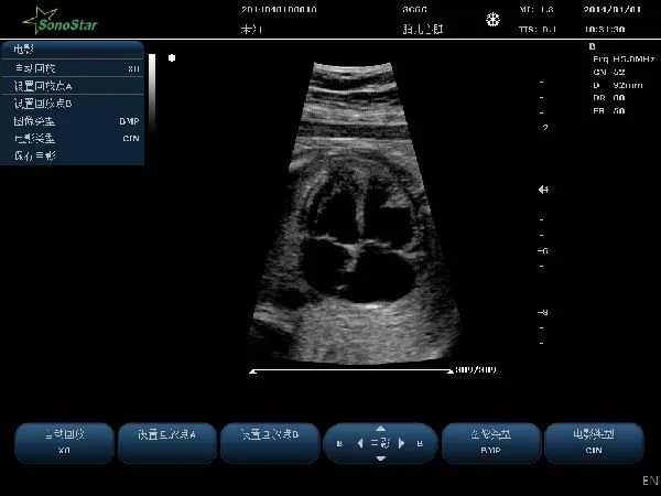
The C10 is a portable colour Doppler system built around a 15-inch or larger medical colour LCD, with two probe sockets as standard. It covers abdominal, gynaecological, obstetric, cardiac, superficial tissue and small parts, nerve, peripheral vascular, cranial, urinary, paediatric, orthopaedic and transrectal examinations, as well as ultrasound-guided interventional procedures.
The main unit measures 400 mm x 394 mm x 172 mm and weighs 7.5 kg without a probe. It starts to the main screen and a live image in 13 seconds or less, runs on mains power or its built-in battery for 2 hours or more of continuous scanning, and stores images on a 128 GB or larger hard disk with report editing and printing on board.
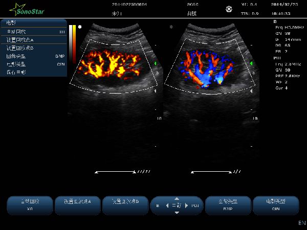
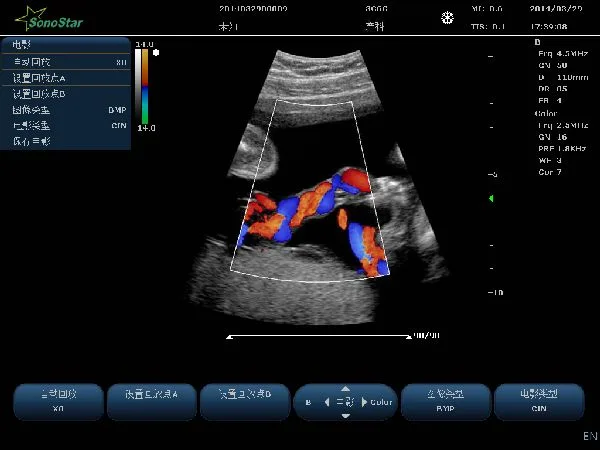
Colour and pulsed-wave Doppler are both available, together with tissue harmonic imaging, spatial compounding and multi-beam formation. Where the department works across several specialties, the nine optional probes cover convex, linear, endocavity, microconvex, rectal and phased array work from a single console.
Technical specifications
Applications and intended use
| Item | Specification |
|---|---|
| Examination areas | Abdomen, gynaecology, obstetrics, cardiac, superficial tissue and small parts, nerve, peripheral vessels, cranial, urinary tract, paediatrics, orthopaedic surgery, transrectal, ultrasound-guided interventional procedures |
| Built-in ECG | 12-lead ECG (optional) |
Main unit and display
| Item | Specification |
|---|---|
| Display | 15-inch or larger high-definition medical colour LCD |
| Probe sockets | 2 or more; 2 fitted as standard |
| Interface languages | English and Chinese (keyboard input, annotations, control panel) |
| Boot time | 13 seconds or less, showing the main screen and image within 13 seconds |
| System upgrade | Supported from the bootloader |
| Safety standard | Meets product safety and quality requirements |
Imaging modes and functions
| Item | Specification |
|---|---|
| Imaging modes | B, M, PW, Color, PDI |
| Display modes | B, dual, quad, B+M, M, B+Color, B+PDI, B+PW, PW, B+Color+PW, B+PDI+PW, B/BC dual real-time |
| Digital imaging techniques | Multi-beam formation, real-time point-by-point dynamic focusing, pulse inversion harmonic imaging, spatial compounding, image enhancement (multi-level) |
| Image zoom | Real-time scanning images (B, B+C, 2B, 4B), continuous zoom |
| Automatic image optimisation | Available in 2D, colour and spectral modes |
| Spectral Doppler | Display and analysis system included |
| Needle guidance | Needle line displayed on B-mode, including a 0-degree needle guide line |
| Aid lines | Lithotripsy line and PICC line displayed on B-mode |
| Presets | Optimal image conditions preset for each organ examined |
2D greyscale parameters
| Item | Specification |
|---|---|
| Greyscale map | 15 steps or more |
| Frame correlation | 8 steps or more |
| Image enhancement | 5 steps or more |
| Spatial compounding | On/off, adjustable |
| Scan density | High, medium, low |
| Image orientation | Up/down and left/right |
| Maximum display depth | 350 mm or more |
| Maximum number of transmit foci | 4 or more |
| Maximum transmit focus positions | 16 or more |
| Maximum B-mode frame rate | 400 frames per second or more |
| TGC segments | 8 or more |
Colour Doppler parameters
| Item | Specification |
|---|---|
| Sample box steering | ±12° or more (linear probe) |
| Doppler frequency | 2 or more bands, visible on screen and independently adjustable |
| Dual display | B/Color dual real-time |
| PRF | 15 steps or more, 0.6-11.7 kHz |
| Colour map | 4 or more |
| Colour correlation | 8 steps or more |
| Post-processing | 4 steps or more |
Spectral Doppler parameters
| Item | Specification |
|---|---|
| Sample volume size and position | Sample volume 1.0-8.0 mm, adjustable |
| PRF | 16 steps or more, 0.7-9.3 kHz, adjustable |
| Sweep speed | 5 steps or more, adjustable |
| Angle correction | -85° to 85°, in 5° steps |
| Spectrum invert | On/off, adjustable |
| Wall filter | 4 steps or more, adjustable |
| Doppler audio | 20 steps or more |
| Automatic spectrum measurement | Supported |
| Blood flow measurement | Supported |
| Steering angle | ±12° or more (linear probe) |
M-mode
| Item | Specification |
|---|---|
| Sweep speed | 5 steps or more |
| Line averaging | 8 steps or more |
Measurement and analysis
| Item | Specification |
|---|---|
| Applicable modes | B-mode, M-mode, spectral Doppler, colour Doppler |
| General measurements | Distance; area and circumference by ellipse and by trace; volume |
| Obstetric and gynaecological | Dedicated gynaecological and obstetric measurement and analysis, including fetal biophysical profile and obstetric formulae for the Chinese population |
| Cardiac function | Supported |
| Peripheral vessels | Blood flow measurement and analysis report |
| Intima-media | Automatic measurement (optional) |
| Doppler measurements | Automatic and manual envelope, with automatic calculation of measured parameters |
Probe specifications
| Item | Specification |
|---|---|
| Frequency | Wideband probes, with independent frequency selection for B/M and for C/D |
| Frequency steps | 5 or more frequencies on convex, linear and phased array probes |
| Needle guide | A needle guide can be specified for every probe type |
Optional probes
| Item | Specification |
|---|---|
| 3C6C | Convex probe, 2.5-6.0 MHz |
| 7L4C | Linear probe, 6.5-10.0 MHz |
| 10L25C | Linear probe, 9.0-14.5 MHz |
| 6E1C | Endocavity probe, 5.0-9.0 MHz |
| 6C15C | Microconvex probe, 5.0-9.0 MHz |
| 6C11C | Microconvex probe, 5.0-9.0 MHz |
| 7I4C | Rectal probe, 6.5-10.0 MHz |
| 2P2F | Phased array probe, 2.0-3.5 MHz |
| 5P2F | Phased array probe, 4.0-6.5 MHz |
Cine loop and storage
| Item | Specification |
|---|---|
| Modes available | All modes |
| Playback | Manual and automatic playback, with adjustable speed |
| B-mode capacity | 5000 frames or more |
| Color/PDI capacity | 2300 frames or more |
| M/PW capacity | 190 s or more |
| Image formats | Single frame: BMP, JPG, FRM; cine: AVI, CIN |
| Offline analysis | FRM and CIN files support offline playback, measurement, parameter adjustment and annotation |
| Hard disk storage | Digital ultrasound image storage of 128 GB or more |
| Integrated workstation | Patient details and the images for that patient can be previewed together |
| One-key storage | A single key press stores the image directly to the hard disk |
| Loop playback | Selected examination images can be played back in a loop |
| USB send and import | Examination data and images can be sent to a USB drive and imported from one |
| Reporting | Report editing and printing, with report templates |
Input and output
| Item | Specification |
|---|---|
| Video output | Composite video, RGB colour video, HDMI |
| Network storage | Data can be transferred to a PC over a network cable |
| DICOM | DICOM 3.0 connection; patient data is uploaded to the DICOM server automatically after the examination |
| USB peripherals | Standard USB foot switch, USB keyboard and mouse, USB barcode scanner |
| FTP transfer | The system can be connected to a PC over a network cable and send examination information to an FTP server |
| Printers | Report printers: monochrome or colour laser, colour inkjet. Video printers: monochrome or colour, digital video |
Power, dimensions and weight
| Item | Specification |
|---|---|
| Battery capacity | 7000 mAh or more |
| Runtime | 2 hours or more of continuous scanning on a full charge |
| Main unit dimensions | 400 mm (length) x 394 mm (width) x 172 mm (depth) |
| Packaging dimensions | 465 mm (length) x 465 mm (width) x 310 mm (height) |
| Main unit weight | 7.5 kg, probe excluded |
| Packaged weight | 12 kg, including the main unit, two probes, power cable, manual, carton and foam |
Specifications updated 2026-11-02
Typical applications
The application scenarios on this page describe the device based on its technical data and configuration. The actual scope of application is the one stated in the product registration certificate. Follow your institution's procedures and the instructions for use.
| Setting / department | Typical use | Configuration note |
|---|---|---|
| Emergency / ICU | Bedside examination of abdomen, heart, pleural and peritoneal fluid | Console with its own colour LCD |
| Obstetrics and gynaecology / urology | Gynaecological, obstetric and urinary tract examinations | Convex and endocavity probes optional |
| Cardiovascular / small parts | Cardiac, peripheral vascular, superficial tissue and small organ examination | Phased array and high-frequency linear probes optional |
| Primary care / health screening | Routine whole-body ultrasound | Multiple probe sockets, configured as needed |
Usage and selection notes
The C10 belongs to the portable colour Doppler series.
The actual scope of application is the one stated in the product registration certificate. Please confirm the model and configuration with our product department.
For contraindications and precautions, refer to the instructions for use. The device must be operated by a licensed physician with the relevant qualifications.
The technical data on this page is taken from the product technical documentation; where it has been updated, the latest version applies. The listed departments and application scenarios describe the device based on its technical data, and the actual scope of application is the one stated in the product registration certificate. This page makes no claim about clinical outcome and does not constitute clinical guidance.
Related products
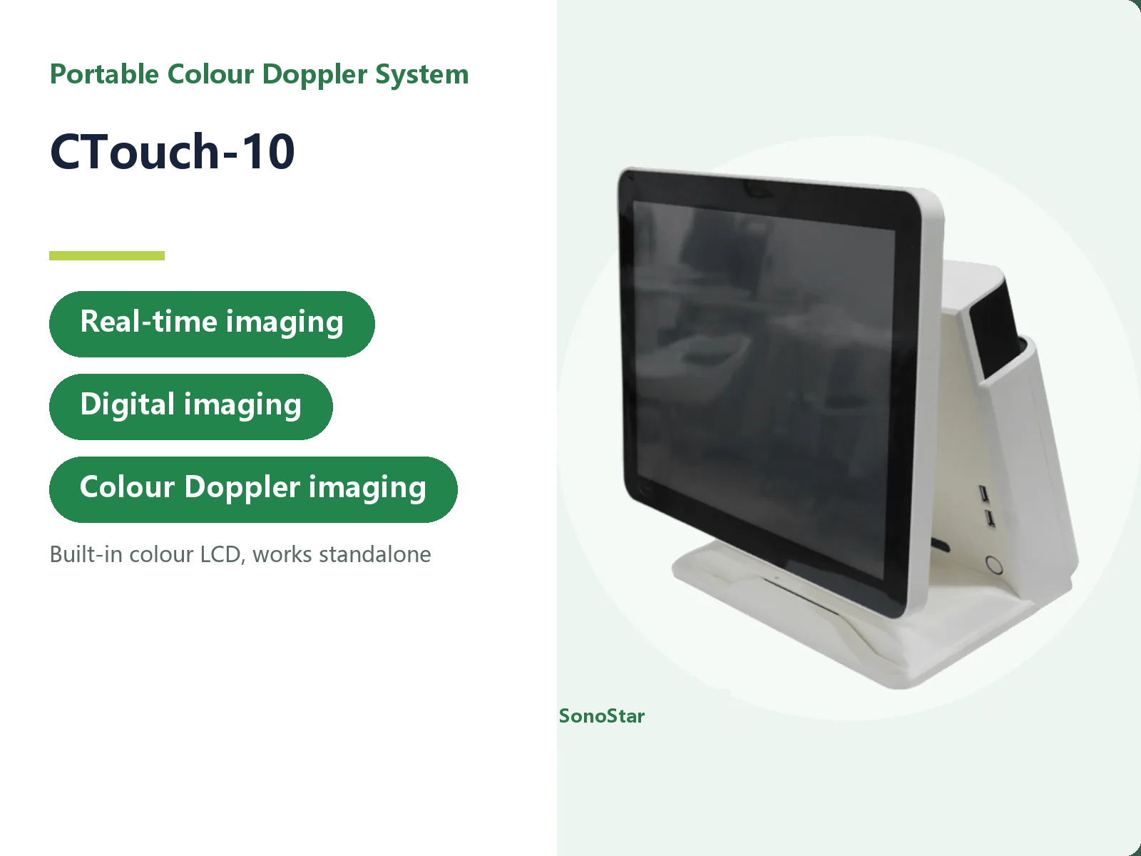
CTouch-10 Portable Colour Doppler Ultrasound System
15-inch touch-screen portable ...
View details →Related articles
Need a quote or a sample for evaluation?
Tell us how you plan to use the system and our sales team will reply within one business day.
Get a quote
