
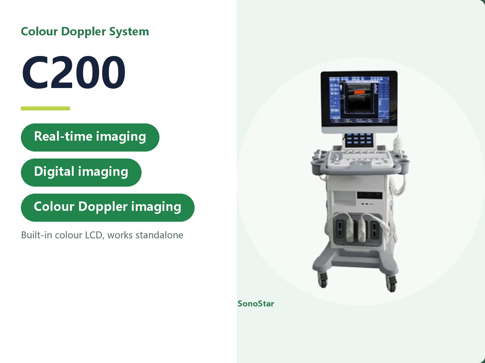
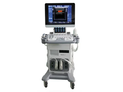
C200 Colour Doppler Ultrasound System
Trolley colour Doppler with a 15-inch touch screen, depth 2-30 cm, frequency 2-10 MHz. Modes B, BB, M, CD, PWD, CFM and THI, optional 3D/4D.
- Brand: SonoStar
- Price: On request
- Probe frequency: Complete system with several probe connectors; a range of probes is available (see specifications)
- Connection: Self-contained colour LCD; works standalone, no external display needed
- Charging: Built-in battery optional (see specifications)
- Category: Trolley Ultrasound
Product overview
The C200 is a trolley-mounted colour Doppler ultrasound system. The main unit and the display sit on a castor base as one body, and the 15-inch colour LCD is a touch screen, so parameter changes and measurements are made directly on the screen without an external display. Plug it in and it works on its own, which suits departments that want a complete system in the room and the freedom to wheel it between rooms.
Imaging modes cover B, BB, M, CD, PWD, CFM, DPDI, HPRF and THI, with real-time B/CD/PW triplex imaging and TGC adjustable by segment. Imaging depth is 2 cm to 30 cm, the frequency range is 2 MHz to 10 MHz, dynamic range is above 120 dB, and zoom runs from 60% to 600% in every mode.
Image processing includes overall gain, 8-segment TGC, M-mode sweep speed, automatic power control and frame correlation, together with brightness and contrast, grey mapping, left-right and up-down flip and rotation, positive and negative display, edge enhancement, noise rejection and speckle reduction. Harmonic imaging cuts speckle noise, and one-key optimisation runs through a full round of image settings automatically.
The body carries four probe connectors, so convex, linear, endocavity, microconvex, rectal or phased array probes can be matched to the department. 3D imaging, a 4D volume probe and its imaging functions are options, and so is the built-in battery. The software includes both human and veterinary modes and switches between them on the same machine. The system can be upgraded and serviced over the network, and reports print through a USB inkjet or laser printer or a video printer.
Typical settings: emergency and ICU for bedside abdomen, cardiac and pleural or peritoneal fluid work; obstetrics, gynaecology and urology with convex and endocavity probes; cardiology and small parts with phased array and high-frequency linear probes; primary care and health screening for routine whole-body scanning. The configuration supplied is the one on the order sheet.
Technical specifications
Key features
| Item | Data |
|---|---|
| Operation | Simple to operate, clean layout |
| Display | 15-inch colour LCD, touch screen |
| Image | High-definition, finely detailed |
| 3D imaging | Optional |
| 4D imaging | 4D probe and imaging functions (optional) |
| Measurement | Wide application and measurement package |
| Storage | Hard disk and USB flash drive |
| Battery | Built-in (optional) |
Main unit and display
| Item | Data |
|---|---|
| Monitor | 15-inch colour LCD with touch screen |
| Imaging modes | B, BB, M, CD, PWD, CFM, DPDI, HPRF, THI; real-time B/CD/PW triplex; TGC gain separately adjustable |
| Imaging depth | 2 cm - 30 cm |
| Frequency range | 2 MHz - 10 MHz |
| Dynamic range | Above 120 dB |
| Zoom | 60% to 600% in every mode |
| Image processing | Overall gain, 8-segment TGC, M-mode sweep speed, automatic power control, frame correlation, brightness and contrast, grey mapping, left-right and up-down flip and rotation, positive and negative display, edge enhancement, noise rejection, speckle reduction |
| Harmonic imaging | Suppresses speckle noise and improves clarity |
| One-key optimisation | Adjusts the image automatically in one step |
| Application software | Human and veterinary software, switchable |
| Software upgrade | Remote upgrade and maintenance over the network |
| Report printing | USB inkjet or laser printer, or video printer |
2D and M-mode
| Item | Data |
|---|---|
| Grey levels | 256 or more |
| Gain | Distance gain compensation, 8 segments or more |
| 2D frame rate | Full field at 18 cm depth, 30 frames per second or more |
| Scanning depth | 30 cm or more |
| Cine memory | 50 frames or more |
| Focus | Manual, visible and adjustable, 4 segments or more |
| Display layouts | B/M, B/D, B/CD, B/D/CD on one screen; M-mode and PWD display size adjustable |
| Zoom | Continuous intelligent zoom on still and moving images |
| Cine playback | Raw-data cine loop; dynamic recording over 10 minutes |
| Compound imaging | Available |
| Presets | User-definable; optimised presets per organ |
Colour Doppler
| Item | Data |
|---|---|
| Colour display | Velocity and power display |
| Colour frame rate | 18 cm depth, 85-degree sector, full colour field, 10 frames per second or more |
| Colour enhancement | Colour Doppler energy map and directional velocity map |
| Region of interest | Linear scan -20 to +20 degrees |
| Display control | Up-down and left-right flip, colour contrast, grey and colour contrast, baseline shift |
| Pseudo-colour | Supported |
Spectral Doppler
| Item | Data |
|---|---|
| Spectrum modes | Pulsed wave Doppler (PWD); continuous wave Doppler (CWD) optional |
| Maximum velocity | PWD unidirectional 6 m/s or more; CWD unidirectional 15 m/s or more |
| Minimum PWD velocity | 5 mm/s or less (non-noise signal) |
| Auto envelope | Automatic envelope measurement and calculation |
| Cine playback | Frozen PWD and CWD traces can be replayed |
| Sample volume | Width and position 0.5-20 mm, stepped adjustment |
| Baseline shift | 6 steps or more |
| Invert display | Yes |
| Angle correction | After Doppler sampling |
| Display control | Invert up-down and left-right, three adjustable scan angles, local zoom (continuous intelligent zoom) |
Probes
| Item | Data |
|---|---|
| Probe connectors | 4 |
| Multi-frequency | Broadband variable-frequency probes; 2D and Doppler can use different frequencies |
| B/D combined | B/PWD, B/CWD, B/CD/PWD triplex |
| Linear probe | Electronic broadband variable frequency, 5.0-12.0 MHz |
| Convex probe | Electronic broadband variable frequency, 2.0-5.0 MHz |
Measurement and analysis
| Item | Data |
|---|---|
| General and dedicated | Distance, length, area, perimeter, volume, angle, stenosis ratio (%), A/B ratio, velocity, pressure gradient (PG), acceleration, resistive index (RI), heart rate, velocity time integral (VTI) |
| Basic measurements | Distance, perimeter, area, slope, heart rate, pressure, residual urine, RI, PI, flow velocity |
| Obstetrics and gynaecology | Gestational age, expected date of delivery, estimated fetal weight, averaging of repeated measurements, S/D ratio |
| Vascular | Vascular measurement, analysis and reporting |
| Spectral analysis | Spectral Doppler analysis software |
| Cardiac and vascular | Ejection fraction (EF), left ventricular volume, cardiac output (CO), pressure half time (PHT), transvalvular gradient, Qp/Qs, velocity integral, body surface area, ICA/CCA diameter ratio, stenosis percentage |
| Custom | Measurement parameters can be edited, added or deleted |
| Packages | Obstetrics and gynaecology, abdominal, urology, endocrine, vascular, cardiac |
Image archive and management
| Item | Data |
|---|---|
| Hard disk | 320 GB built-in, real-time storage of still and moving images |
| On-screen clipboard | One key stores still and moving images to a side clipboard for later comparison |
| Offline review | Brightness, contrast, Doppler baseline and sample angle can be adjusted on recalled images and saved as AVI, TIF or BMP |
| Interfaces | USB, S-VIDEO, VGA, network image transfer |
| Network | Real-time image exchange, storage and playback over the network |
| File formats | AVI, JPG, BMP, PNG, TIF, DCM; stored and managed on the hard disk, a USB drive or optical media |
Configuration
| Item | Data |
|---|---|
| Standard | Main unit x1, convex probe x1, power adapter and accessories x1 set |
| Optional | Linear, endocavity, microconvex, rectal and phased array probes; 3D imaging; 4D volume probe and imaging; laser printer; video printer |
Specifications updated 2026-11-01
Typical applications
The application scenarios on this page describe the device based on its technical data and configuration. The actual scope of application is the one stated in the product registration certificate. Follow your institution's procedures and the instructions for use.
| Setting / department | Typical use | Configuration note |
|---|---|---|
| Emergency / ICU | Bedside scanning of the abdomen, the heart and pleural or peritoneal fluid | Self-contained unit with a colour LCD |
| Obstetrics / gynaecology / urology | Gynaecological, obstetric and urinary tract examinations | Convex and endocavity probes available |
| Cardiology / small parts | Cardiac, peripheral vascular, superficial tissue and small organ examinations | Phased array and high-frequency linear probes available |
| Primary care / health screening | General whole-body ultrasound | Several probe connectors, configured to need |
Usage and selection notes
The C200 is a complete-system product and belongs to a different product line from the wireless probes.
A number of probe types can be fitted, so the configuration supplied is the one on the order sheet. Please confirm the selection with our product department.
For contraindications and precautions, refer to the instructions for use. The device must be operated by a licensed physician with the relevant qualifications.
The technical data on this page is taken from the product technical documentation; where it has been updated, the latest version applies. The listed departments and application scenarios describe the device based on its technical data, and the actual scope of application is the one stated in the product registration certificate. This page makes no claim about clinical outcome and does not constitute clinical guidance.
Related products
Related articles
Need a quote or a sample for evaluation?
Tell us how you plan to use the system and our sales team will reply within one business day.
Get a quote
