
Handheld Ultrasound
The probe is the system. The ultrasound engine, the battery and the wireless module all sit inside the probe handle, and the image travels over the probe's own Wi-Fi straight to a phone, tablet or computer — pick up the probe and start scanning, whether you are at the bedside, in an ambulance or in a village clinic.
Selection guide · how to choose by setting (click to expand)
Clinical departments
Anaesthesia, pain management, nursing and line placement (PICC / CVC), nephrology, vascular surgery, thyroid and breast surgery, rehabilitation, orthopaedics and traumatology, acupotomy and acupuncture, critical care, emergency medicine, pre-hospital care, respiratory medicine, cardiology, gastroenterology, neurology, hepatobiliary and general surgery, urology, gynaecology, obstetrics, reproductive medicine and neonatology — the bedside needs of 22 clinical departments, met with one probe.
Examination areas
From 10 mm at the surface to 305 mm deep, each depth has its matching probe:
- Superficial 20-100 mm | high-frequency linear 7.5–14.0 MHz: thyroid, breast, musculoskeletal, nerve, vascular, carotid, paediatric and small-parts work.
- Abdominal and deep 90-305 mm | convex 3.2–5.0 MHz: liver, gallbladder, spleen, kidney, gynaecology, obstetrics, urinary tract, lung.
- Cardiac | phased array 2.2–3.6 MHz: assessment of cardiac structure and blood flow.
- Endocavity 30-120 mm | 6.6 MHz / 8 MHz: transvaginal, transrectal and prostate examinations.
- Small body 60-90-120-160 mm | micro-convex 5.0–7.0 MHz: neonatal and paediatric abdomen.
- Ophthalmic: uses the dedicated ophthalmic probe; see the Ophthalmic Ultrasound section.
Device types and probe configurations
41 models and 9 transducer types, covering 10 mm through 305 mm from one supplier.
| Transducer type | Representative models | Frequency | Depth | Main applications |
|---|---|---|---|---|
| Linear / high-frequency linear | 5L, 5P, 6L, 7L | 7.5–10.0 MHz / 10.0–14.0 MHz | 20-100 mm | Superficial, vascular, nerve, musculoskeletal, thyroid and breast |
| Ultra-high-frequency linear | 7H, 8H, 8S, 9H, 9S | 16.0–20.0 MHz / 18.0–24.0 MHz | 10-40 mm | Fine structures closer to the surface |
| Convex | 5C, 6C, 7C, 8C, 9C | 3.2–5.0 MHz | 90-305 mm | Abdomen, gynaecology, obstetrics, urinary |
| Micro-convex | 5MN, 5ML | Micro-convex 5.0–7.0 MHz | 60-90-120-160 mm | Neonates, children, small patients |
| Phased array / cardiac | 5PA, 6PA, 5CPL in cardiac mode | 2.2–3.6 MHz / 3.2–5.0 MHz | 90-240 mm | Cardiac |
| Endocavity | 5T, 5CT in endocavity mode | 6.6 MHz / 8 MHz | 30-120 mm | Transvaginal, transrectal, prostate |
| Biplanar rectal | 5CR, 9CR | 6.5 MHz / 8 MHz | 40-60-80-100 mm | Transrectal |
| Dual-head / three-in-one | 5CP, 5CL, 5CPL, 6CPL, 7CPL, 8CPL, 9CPL | Convex + linear (+ cardiac) | Combined coverage | One probe covers several examination types |
| Special-purpose | 6F with finger sleeve, 6X with magnetic navigation | 7.5–10.0 MHz | 20-100 mm | Specific procedural settings |
Key specifications
- Imaging modes: B, B+M, Color (colour Doppler), PDI (power Doppler), PW (pulsed-wave Doppler)
- Gain 30–105 dB; noise reduction levels 0-1-2-3-4; dynamic range 40-50-60-70-80-90-100-110
- Needle guide lines: in-plane and out-of-plane modes
- Image transmission: the probe carries its own Wi-Fi signal and does not depend on a network being available on site — it works where there is no coverage
- Battery: about 3 hours of scanning, about 2-3 hours to charge; wireless or wired charging, and the 9 series has a replaceable battery
- Measurements: length, angle, area/circumference, trace, area ratio; obstetrics adds crown-rump length, biparietal diameter, gestational sac diameter, femur length, head circumference, abdominal circumference and estimated fetal weight
Where it fits
- Primary care facilities (township health centres, community health centres, village clinics): routine examination of abdominal organs, carotid, thyroid, breast, uterus and adnexa, prostate and superficial masses.
- Private and specialist clinics, health-screening centres: mobile routine screening.
- Hospital clinical departments (anaesthesia, pain, emergency, critical care, respiratory, nephrology, vascular surgery and others): bedside assessment and visual guidance.
- Mobile and follow-up settings (ambulances, field and temporary clinics, home visits, care homes, postnatal centres).
How it differs from our other ultrasound ranges
Our three ultrasound ranges are divided by where they are used, and they are not interchangeable:
- This range — the probe is the system and there is no separate main unit: bedside work, mobile use, limited space.
- Portable Ultrasound — with its own display: outpatient rooms, wards and house calls, where a larger screen and full workstation functions are needed.
- Trolley Ultrasound — a cart-mounted system: routine examinations in a fixed department.
Selection starts with two questions: where will it mainly be used, and does it need to move at a moment's notice?
Common purchasing questions
Q: Can it be used where there is no Wi-Fi, such as an ambulance or a field setting?
A: Yes. The probe generates its own Wi-Fi, so the working environment does not need a network.
Q: Do I have to choose the colour version?
A: It depends on the work. For needle and line guidance, where the task is to see the needle tip and its position, the greyscale (B / B+M) version is sufficient. Where blood flow has to be assessed — vascular work or cardiac — choose a configuration with Color / PDI / PW.
Q: How long does the battery last, and how is it charged?
A: About 3 hours of continuous scanning, about 2-3 hours to charge; a USB cable, a wireless charger or a power bank can all top it up, and some models accept a replacement battery.
Q: Can it produce reports and print images? What accessories and consumables go with it?
A: The device is positioned for emergency use, initial screening and visual guidance, and has no built-in reporting function; images can be exported to a computer for editing or sent to a printer. Common accessories are ultrasound carts, wireless charging pads, needle guides and carry bags. The routine consumable is coupling gel, and the single-use needle guides used for interventional guidance are consumables — see Probes & Accessories.
The information on this page is a technical reference based on product specifications; the applicable scope is that stated in the product registration certificate. Contraindications and precautions are set out in the product manual, and the device must be operated by a licensed physician holding the relevant qualifications. Nothing on this page constitutes a claim of clinical efficacy.
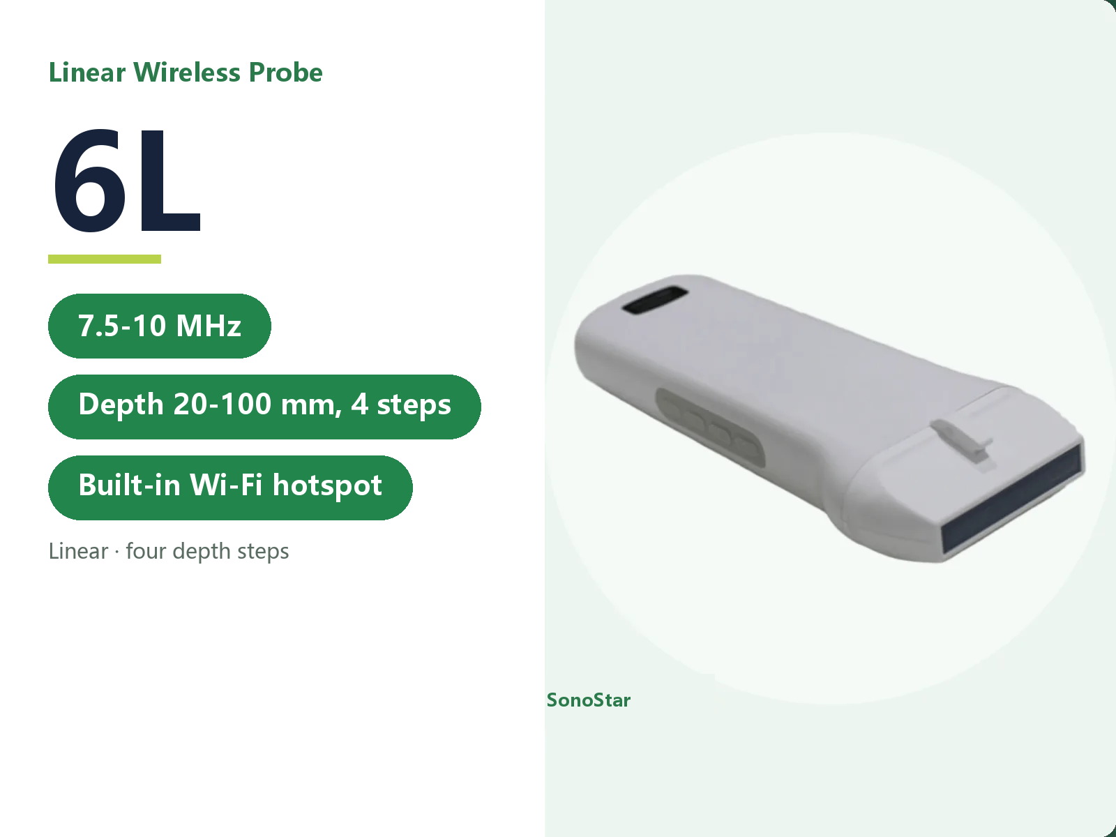
6L Linear Wireless Ultrasound Probe
7.5-10.0 MHz, depth 20-100 mm in four steps, covering both superficial and slightly deeper sites
View details →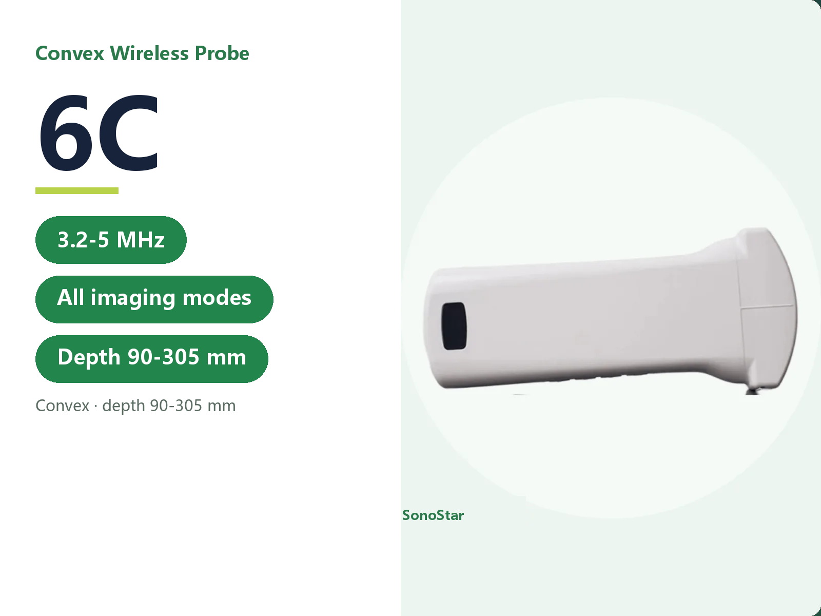
6C Convex Wireless Ultrasound Probe
3.2-5.0 MHz convex, full imaging modes, depth 90-305 mm
View details →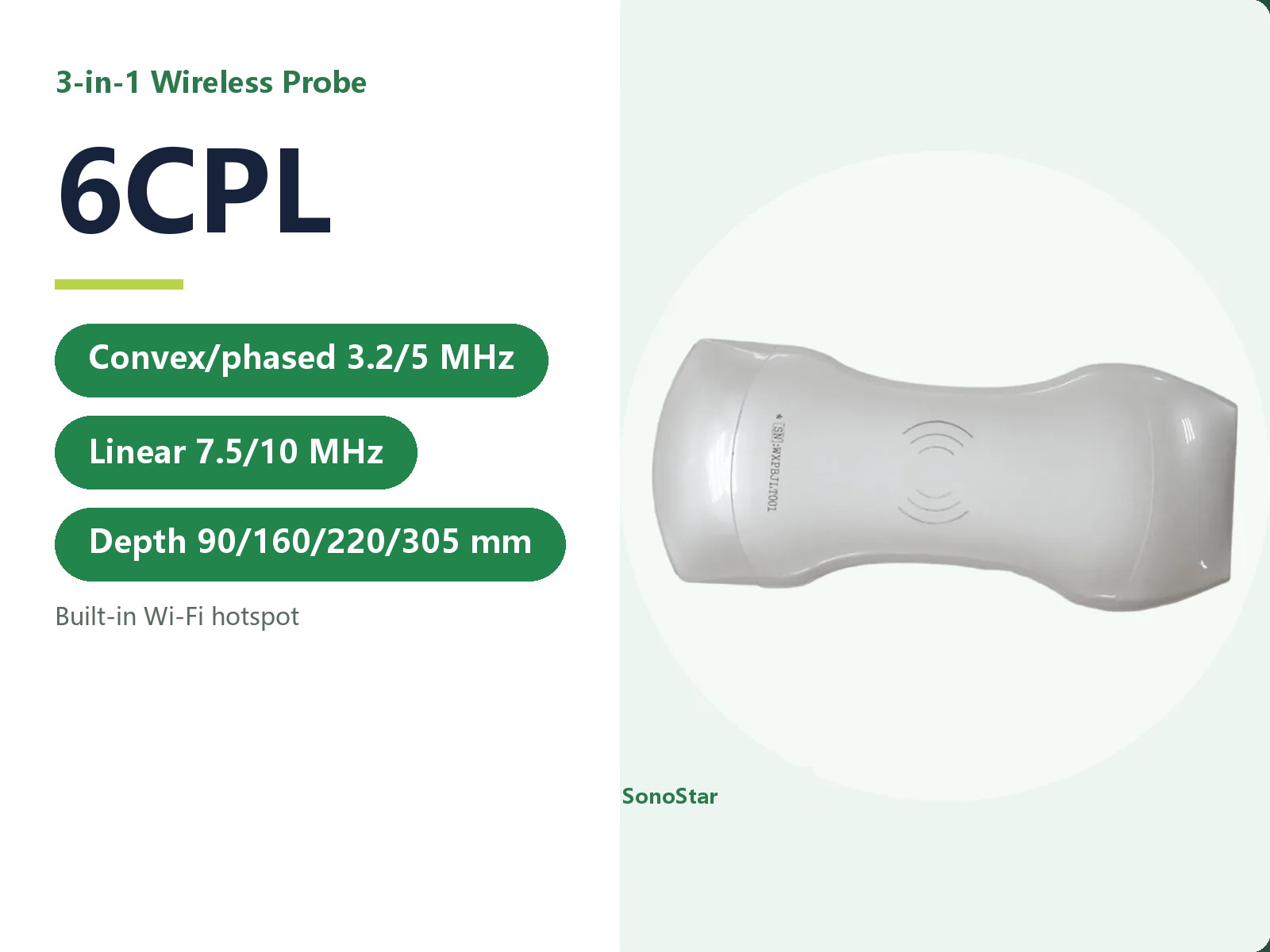
6CPL Three-in-One Wireless Ultrasound Probe
Convex and phased array 3.2-5.0 MHz, linear 7.5-10.0 MHz, cardiac mode 3.2-5.0 MHz, full imaging modes
View details →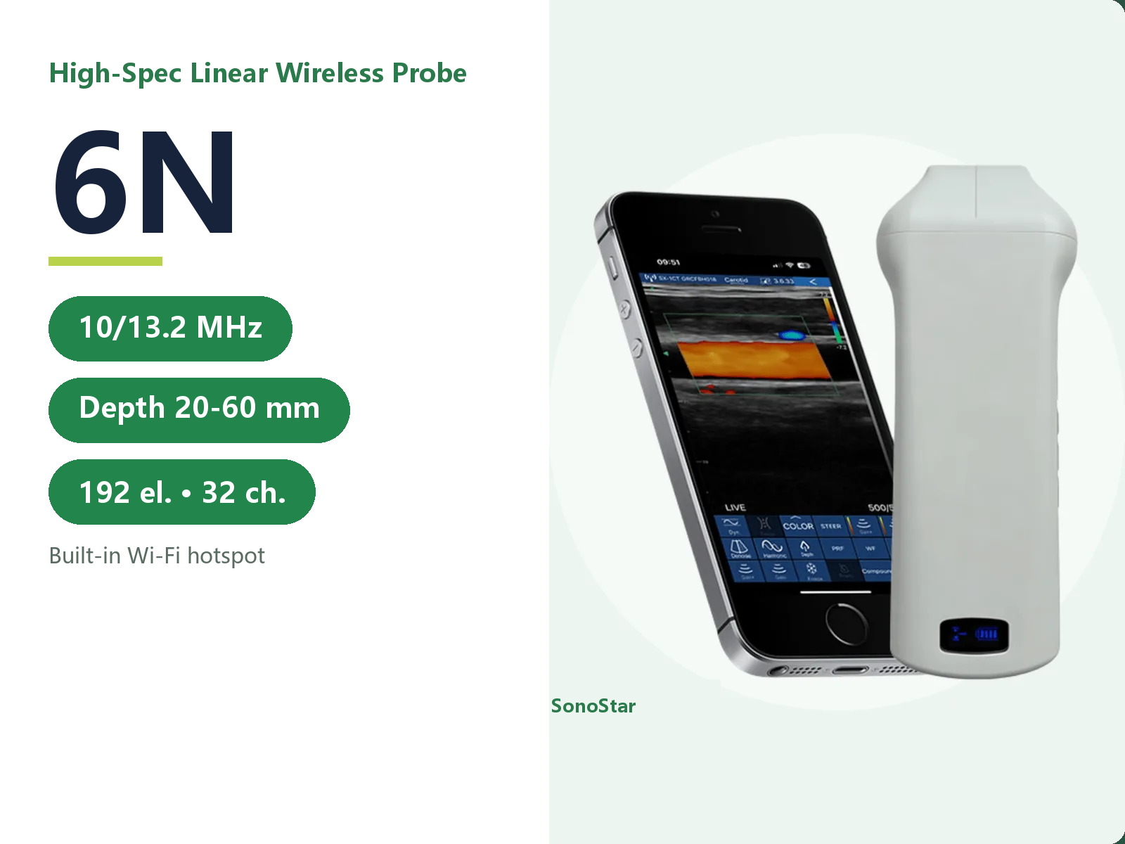
6N High-Configuration Linear Wireless Ultrasound Probe
10.0-13.2 MHz linear, full imaging modes, depth 20-60 mm
View details →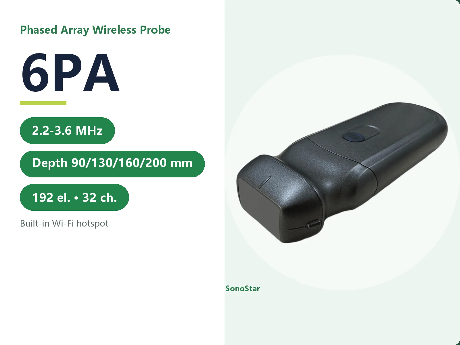
6PA Phased Array Wireless Ultrasound Probe
2.2-3.6 MHz phased array, full imaging modes including CW, depth 90/130/160/200 mm
View details →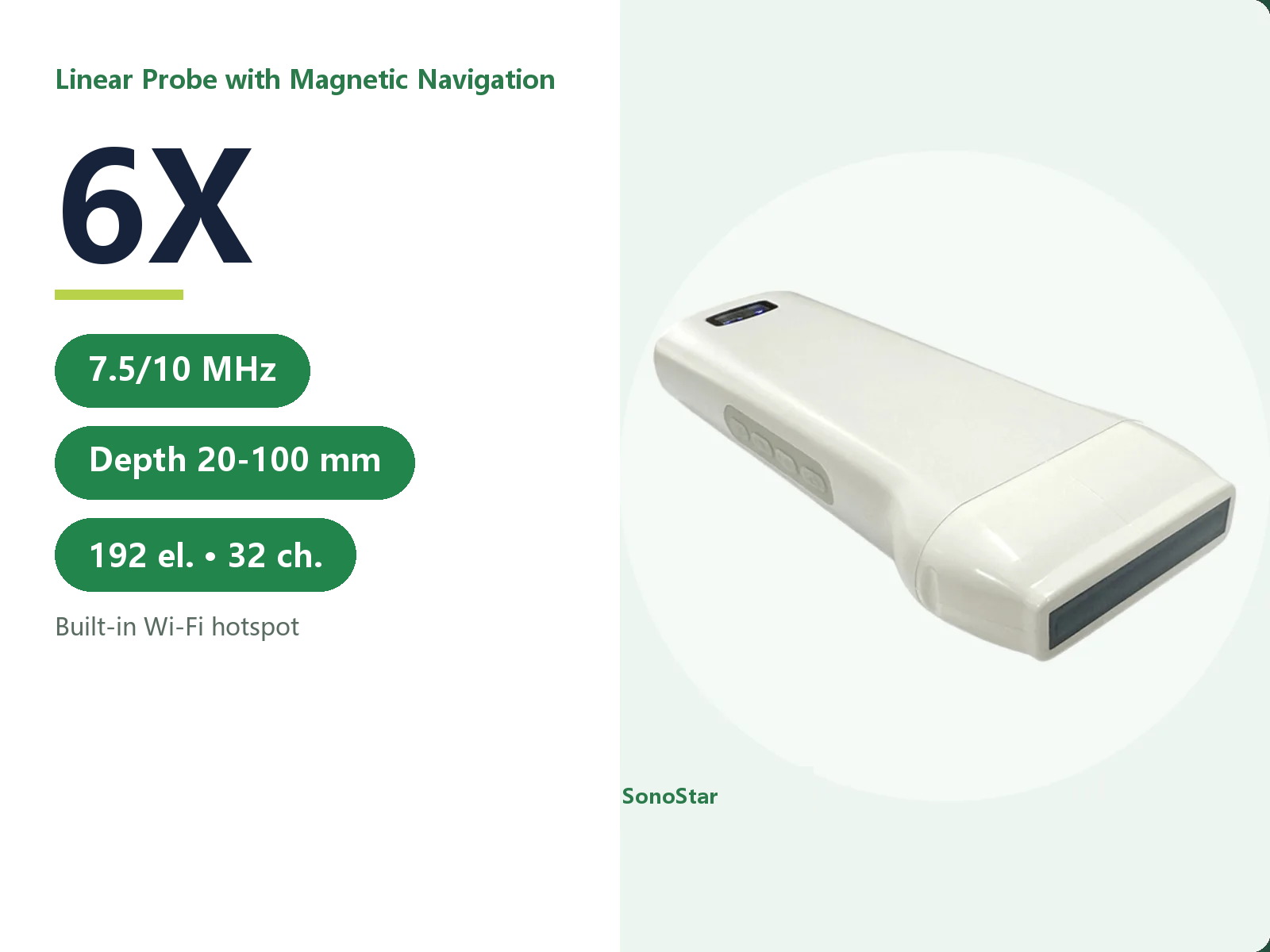
6X Linear Wireless Ultrasound Probe with Magnetic Navigation
7.5-10.0 MHz linear with magnetic needle navigation, full imaging modes, depth 20-100 mm
View details →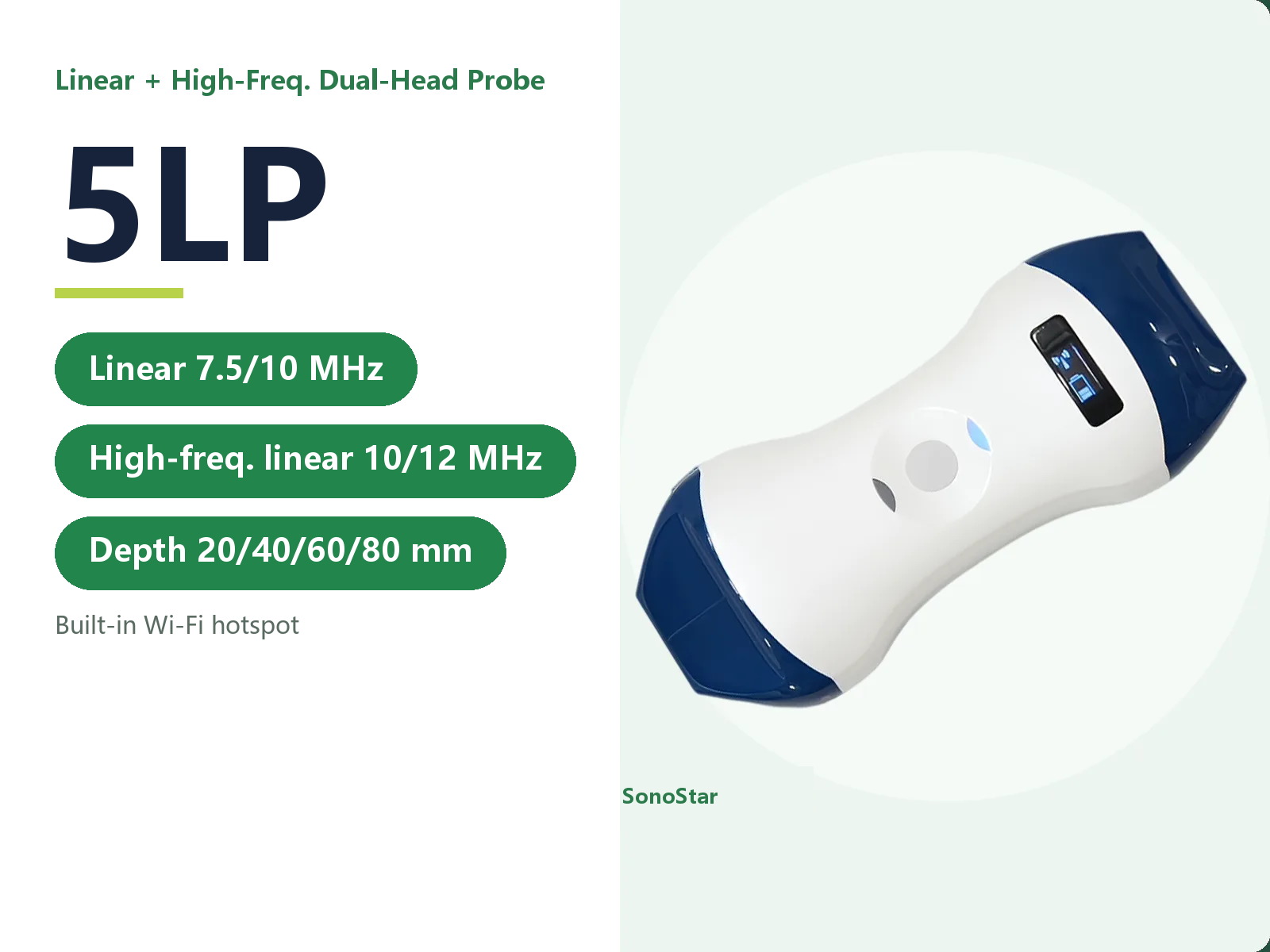
5LP Linear + High-Frequency Linear Dual-Head Wireless Probe
Linear 7.5-10.0 MHz plus high-frequency linear 10.0-12.0 MHz, full imaging modes
View details →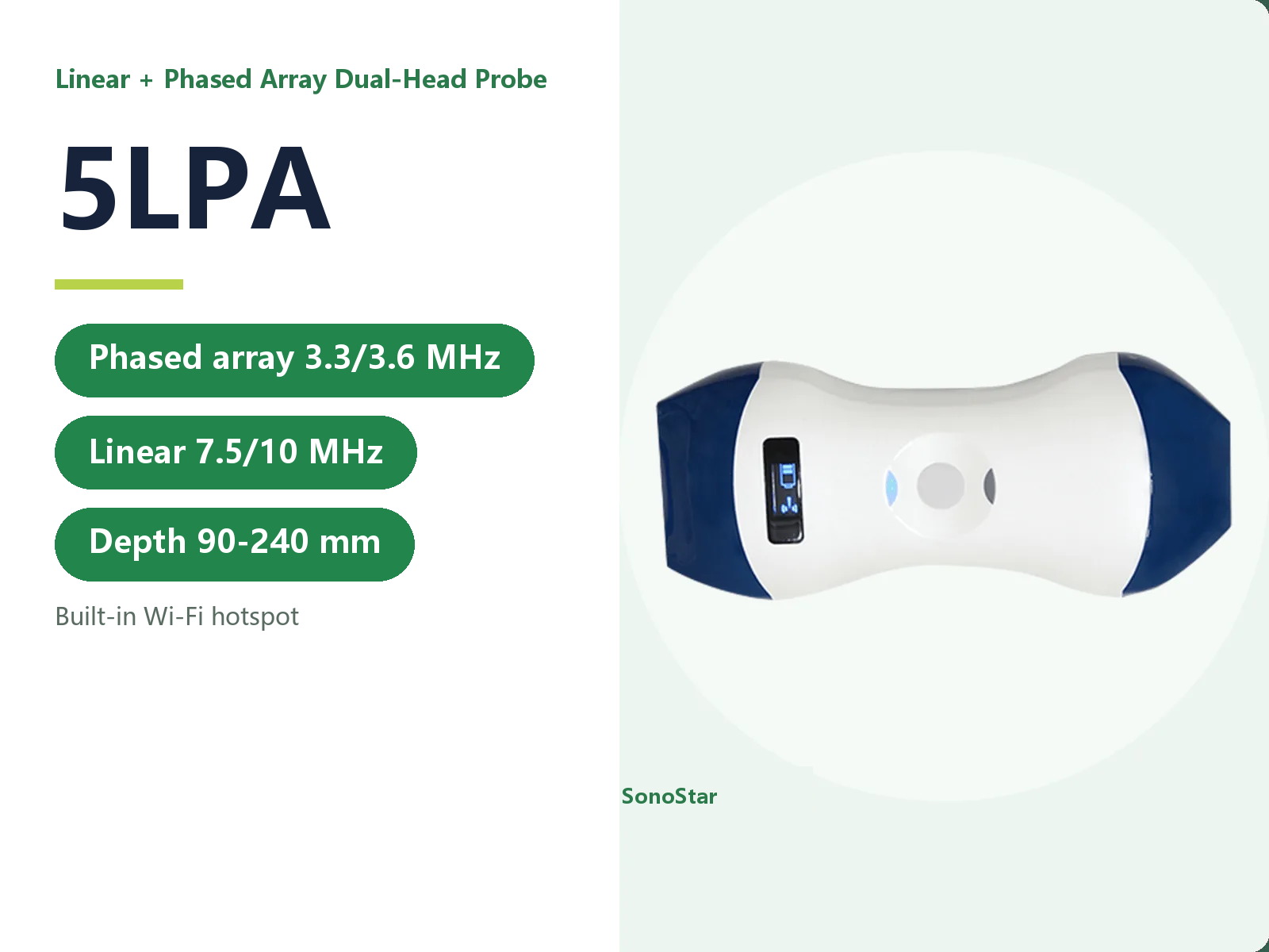
5LPA Linear + Phased Array Dual-Head Wireless Probe
Phased array 3.3-3.6 MHz plus linear 7.5-10.0 MHz, depth to 240 mm
View details →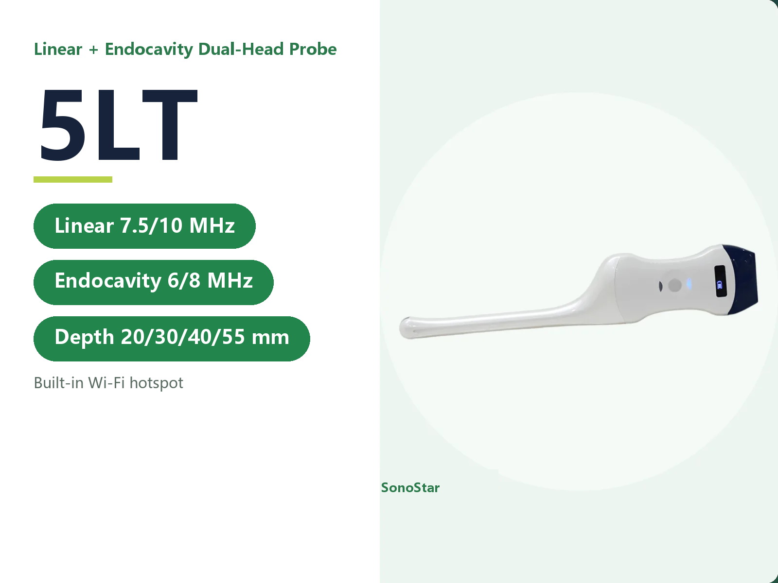
5LT Linear + Endocavity Dual-Head Wireless Probe
Linear 7.5-10.0 MHz plus endocavity 6.0-8.0 MHz, full imaging modes
View details →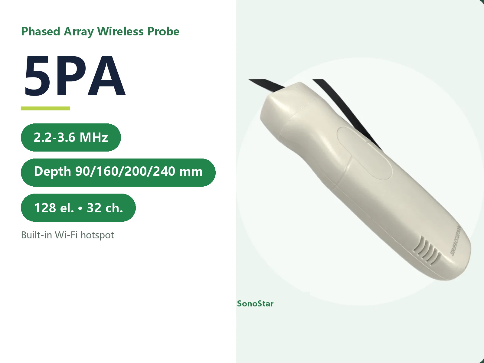
5PA Phased Array Wireless Ultrasound Probe
2.2-3.6 MHz phased array, full imaging modes, depth 90/160/200/240 mm
View details →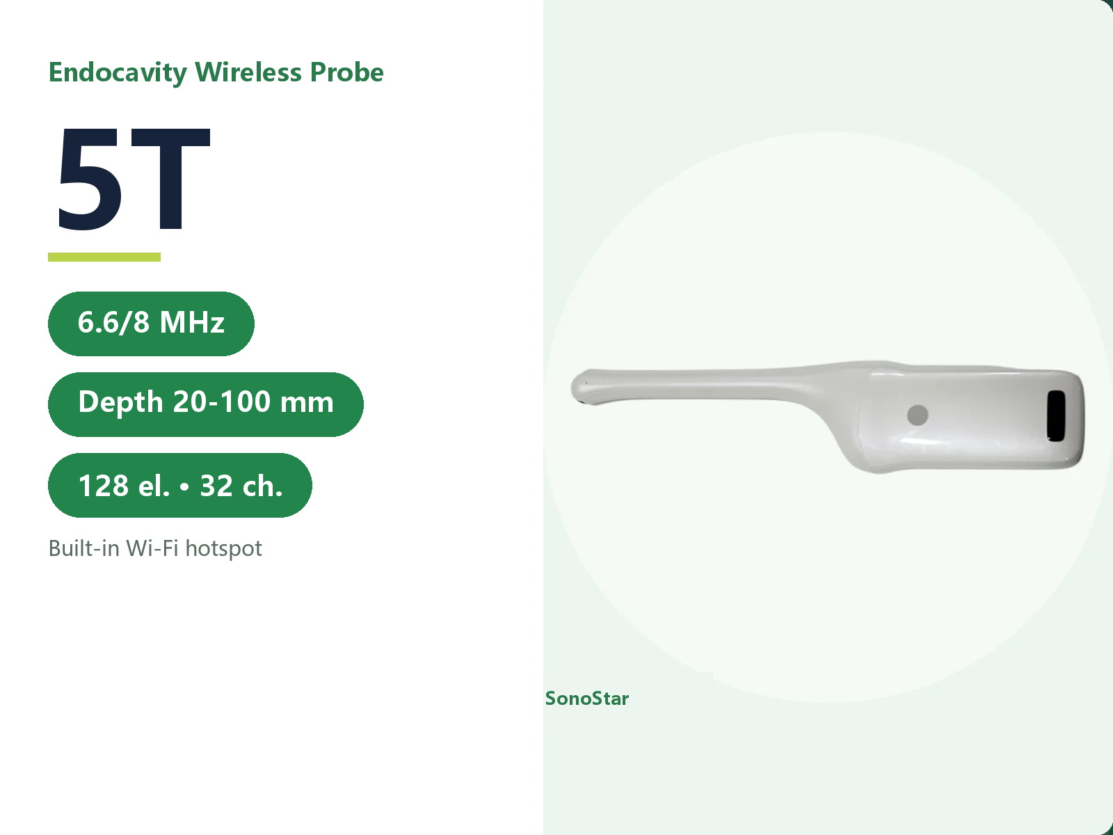
5T Endocavity Wireless Ultrasound Probe
6.6-8.0 MHz endocavity, full imaging modes, depth 20-100 mm
View details →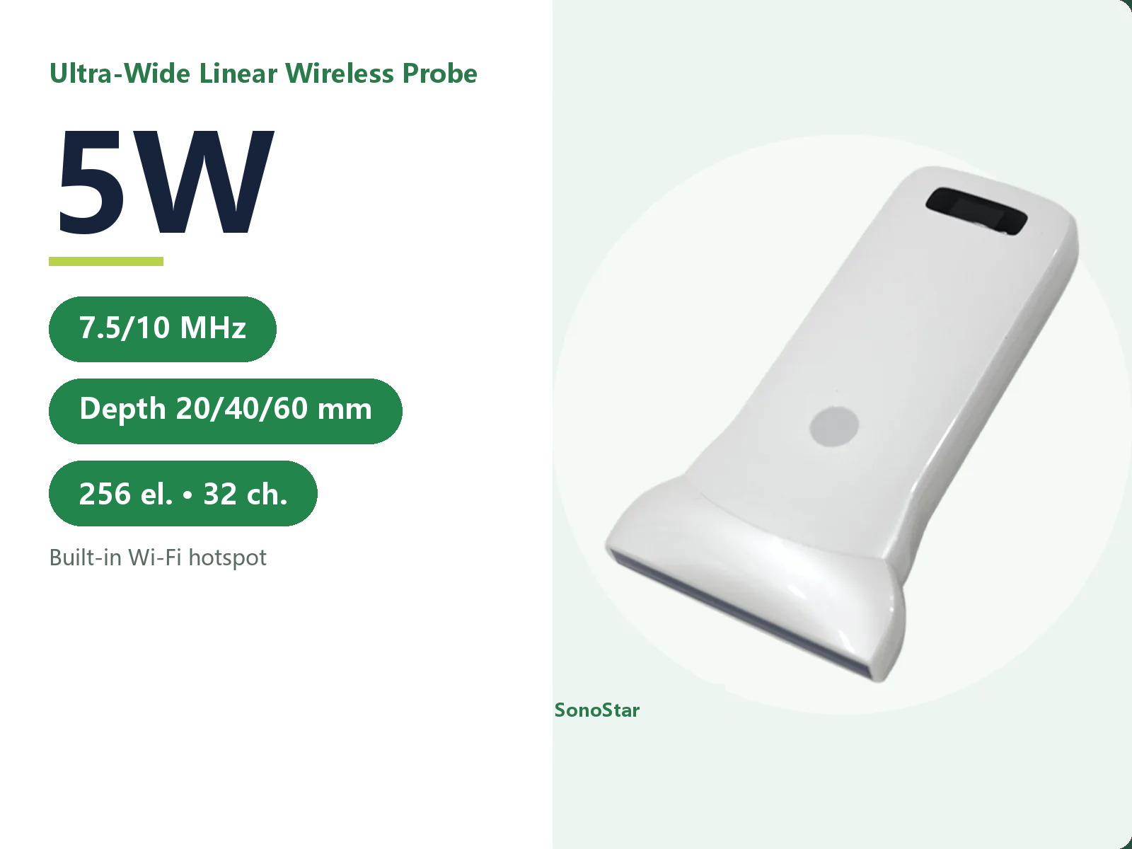
5W Ultra-Wide Linear Wireless Ultrasound Probe
7.5-10.0 MHz ultra-high-frequency linear with an 80 mm face and wide field of view
View details →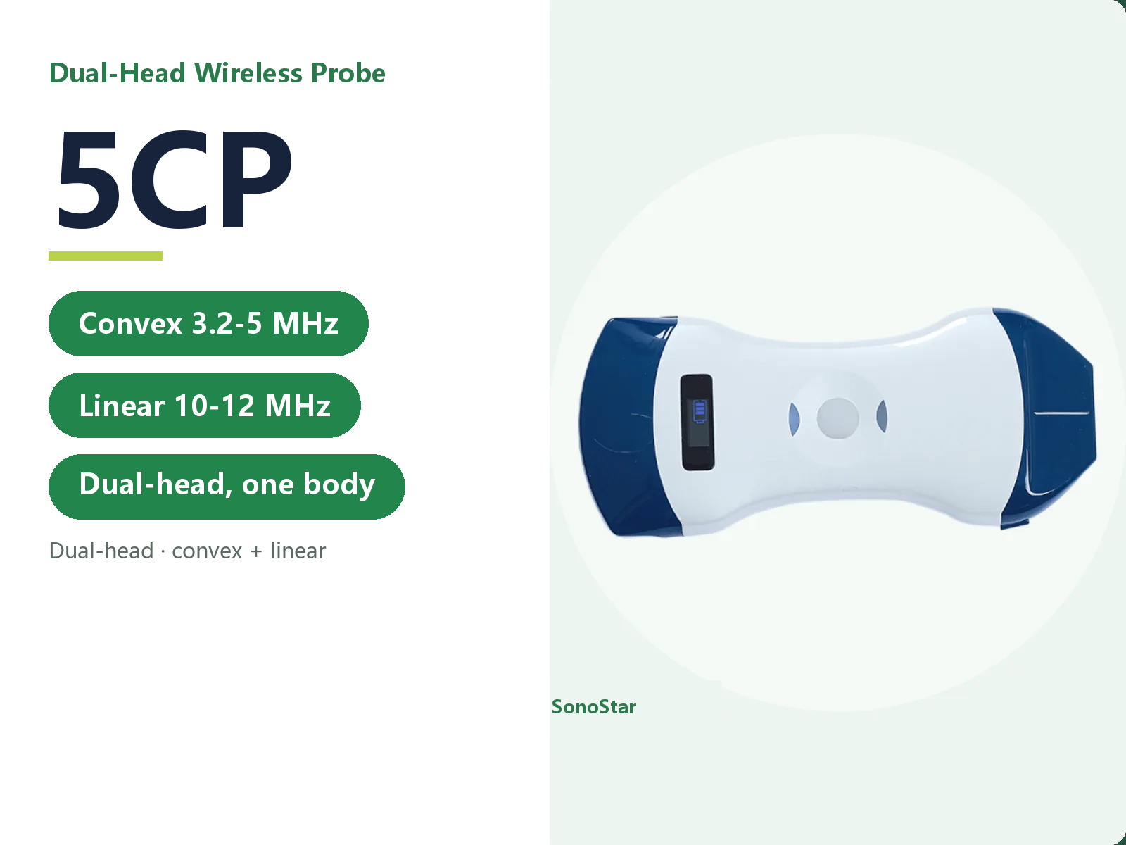
5CP Dual-Head Wireless Ultrasound Probe (Convex + Linear)
Convex and linear heads in one probe - two examination types without changing probe
View details →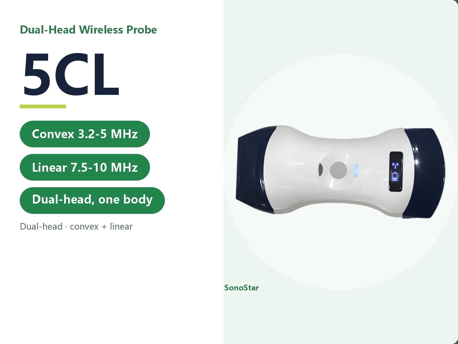
5CL Dual-Head Wireless Ultrasound Probe (Convex + Linear)
Convex and linear heads in one colour Doppler probe, abdomen and superficial covered at once
View details →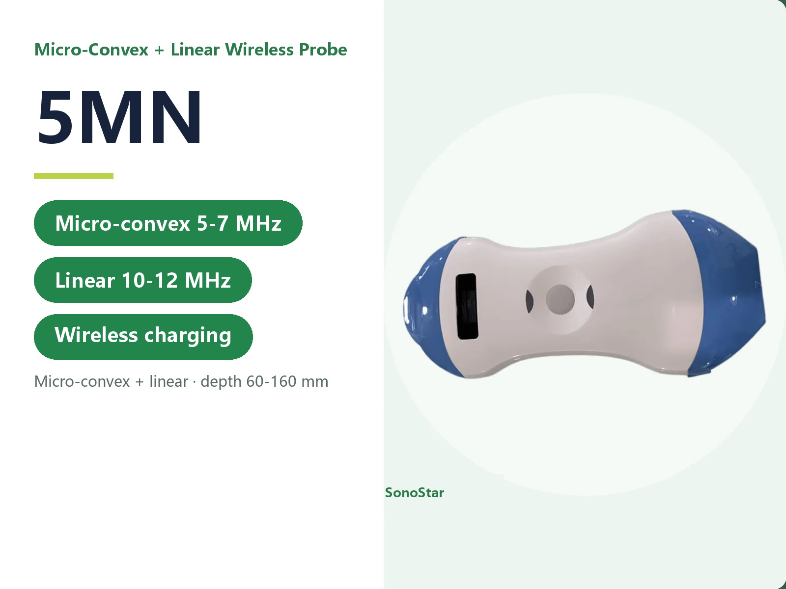
5MN Micro-Convex + Linear Wireless Ultrasound Probe
Micro-convex plus linear, wireless charging, covering paediatric abdomen and superficial work
View details →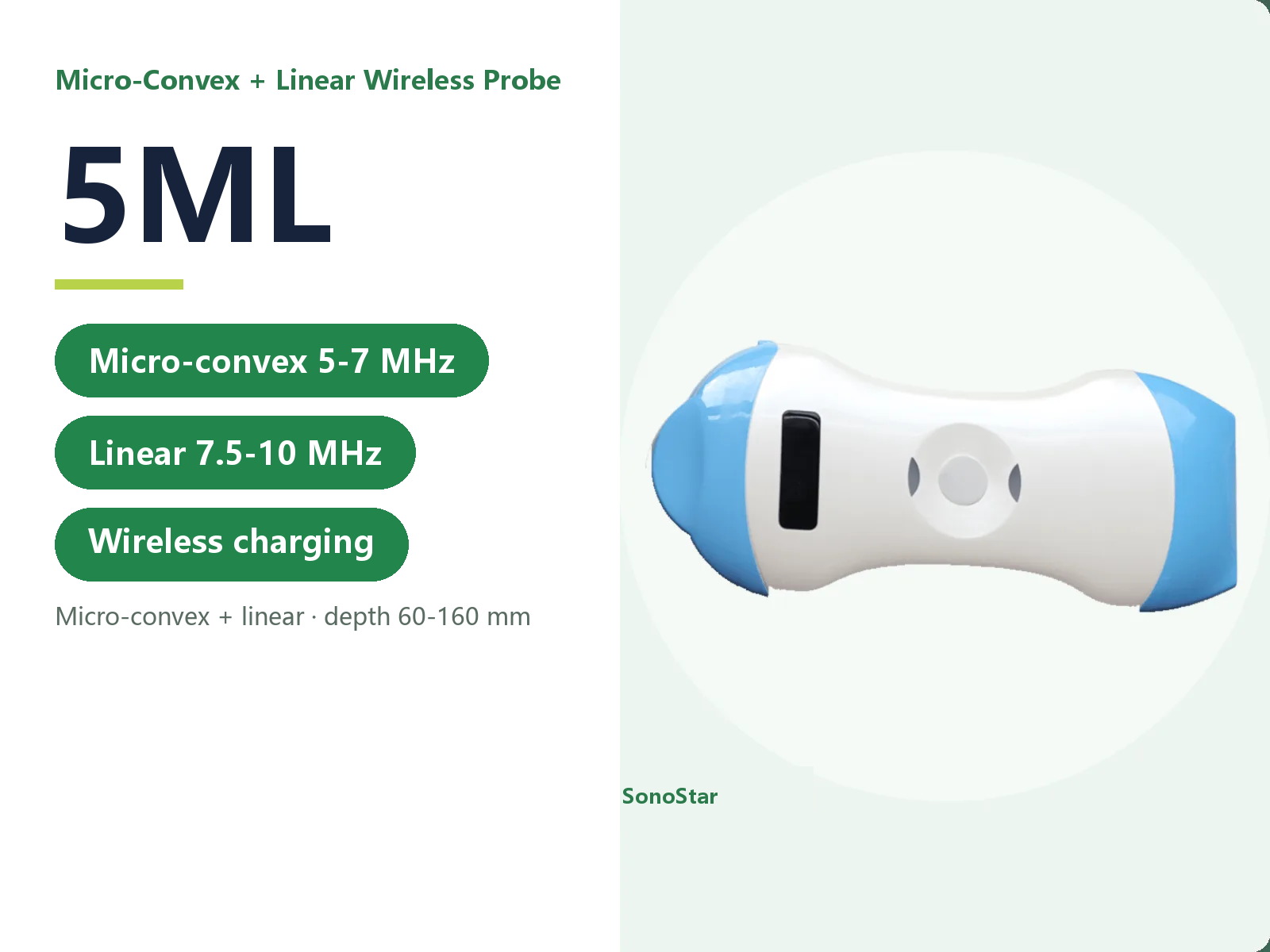
5ML Micro-Convex + Linear Wireless Ultrasound Probe
Micro-convex plus linear at 7.5-10.0 MHz, suited to small patients
View details →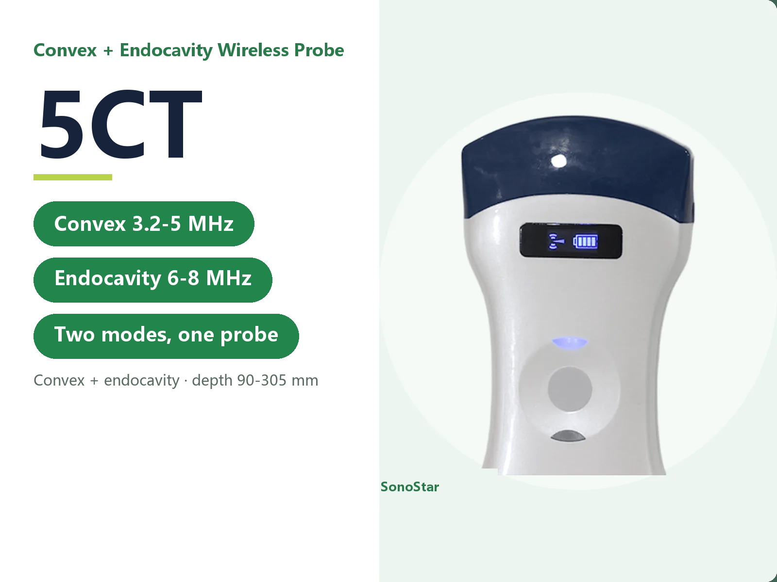
5CT Convex + Endocavity Wireless Ultrasound Probe
Convex and endocavity modes in one probe, abdominal and intracavity work covered together
View details →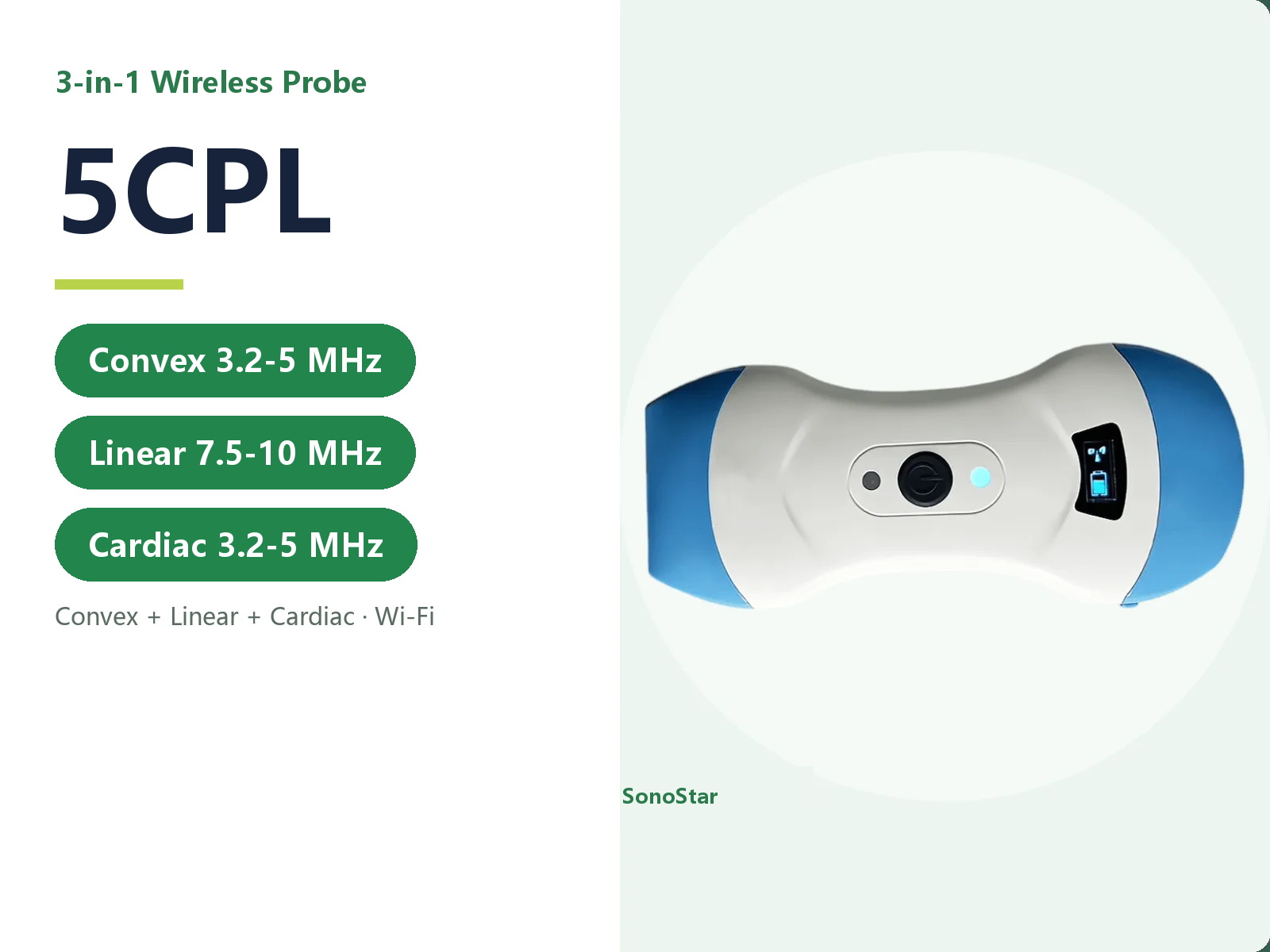
5CPL Three-in-One Wireless Ultrasound Probe (Convex + Linear + Cardiac)
Convex, linear and cardiac in one probe - three examination types from the first unit
View details →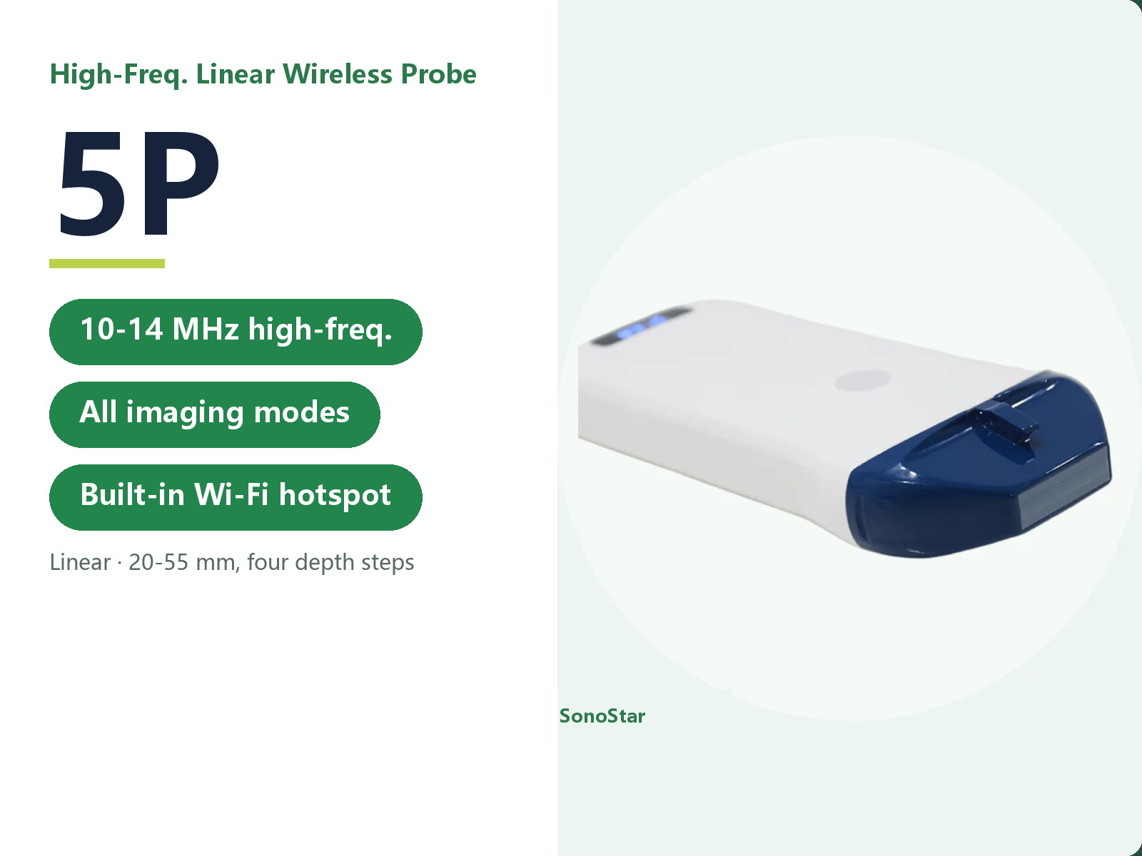
5P High-Frequency Linear Wireless Ultrasound Probe
10.0-14.0 MHz high-frequency linear, the usual choice for superficial needle guidance
View details →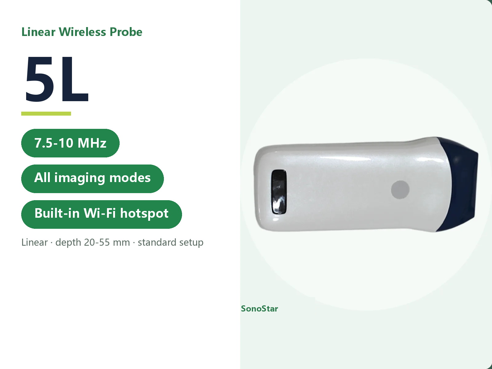
5L Linear Wireless Ultrasound Probe
7.5-10.0 MHz linear, the routine superficial configuration with 20-55 mm depth
View details →Frequently asked questions
Common questions about products and selection
Does the handheld scanner need an external main unit?
No. The ultrasound engine, battery and wireless module are all built into the probe handle, so the probe alone is enough to start scanning.
Where is the image displayed?
The probe's built-in Wi-Fi sends the image straight to a smartphone, tablet or computer, with no cable between them.
Which settings is it suited to?
Mobile settings such as the bedside, ambulances and small primary-care clinics. It is ready once switched on and easy to carry.
Which clinical departments use it?
Bedside examination in anaesthesia, pain management, nursing and vascular access (PICC/CVC), nephrology, vascular surgery, thyroid and breast surgery, rehabilitation, orthopaedics, critical care, emergency, obstetrics and gynaecology, and urology, among others.
How do I choose a probe?
Select by examination depth, from 10 mm at the surface to 305 mm deep. If you are unsure which model fits, tell us the site you will be scanning and we will help you match it.
Didn't find what you need?
Tell us how you will use the system, and we will propose an ultrasound configuration to match.
Contact us
